You're hurt. You're overwhelmed.
Your recovery starts here.
Expert personal injury chiropractic that protects your health and your case. Movement-based recovery backed by precise diagnosis and defensible documentation.
Our Framework
Five steps to full recovery
A systematic approach to diagnosing, documenting, and treating personal injury — so nothing gets missed.
Comprehensive Evaluation
Orthopedic, neurological, and functional examination including cranial nerve assessment most providers skip.
Precise Diagnosis
Advanced imaging interpretation and differential diagnosis. We identify injuries others miss — radiculopathy, ligament instability, mild TBI indicators — and document them defensibly.
Movement-Based Rehabilitation
Progressive resistance, vibration therapy, and targeted manual care. We rebuild function, not just reduce pain. Every session is documented with measurable outcomes.
Defensible Documentation
Reports built for attorneys, adjusters, and courtrooms. Impairment ratings, causation analysis, and treatment necessity — all in one package.
Maximum Medical Improvement
We don't stop until you've reached your best possible outcome. Final reports include permanent impairment ratings and future care recommendations when warranted.
Treatment Methods
Rehabilitation that rebuilds function
Three evidence-based modalities, each targeting a different dimension of injury recovery.
Progressive Resistance Training
Rebuilds movement patterns damaged by injury. Progressively restores strength, coordination, and endurance along proper biomechanical lines of force.
Functional Resistance RehabilitationVibration Therapy
Restores stability and neuromuscular coordination. Whole-body vibration activates the neuromuscular system, retrains proprioception, balance, and deep stabilizer function.
Neuromuscular Activation PlatformFocused Shockwave Therapy
For injuries that resist standard treatment. Acoustic energy targeted at damaged tissue triggers cellular repair. Positioned as our escalation tool — we don't give up.
Advanced Tissue RecoveryFor Personal Injury Attorneys
Documentation built for negotiation
We understand what your cases need. Every report is designed to support demand packages, mediations, and trial.
- ✓ Complete injury reports with mechanism of injury analysis
- ✓ AMA Guides impairment ratings (5th & 6th edition)
- ✓ Cranial nerve and neurological examination findings
- ✓ Causation opinions linking injuries to collision
- ✓ Future care recommendations and cost projections
- ✓ 100% lien-based — zero cost to your client upfront

Our Philosophy
Movement is medicine
Injuries heal with guided movement, not rest. We restore function through progressive, measured rehabilitation — backed by evidence and documented for your case.
-
🧬
Tissue Remodeling
Controlled loading stimulates collagen alignment and tissue repair along functional lines of force.
-
🧠
Neuroplastic Recovery
Movement retrains disrupted motor patterns and restores proprioceptive accuracy after injury.
-
📊
Measurable Progress
Every session produces objective data — range of motion, strength testing, functional capacity — for your records and your case.
-
🛡️
Defensible Treatment
Evidence-based protocols with documented medical necessity at every step. Built to withstand scrutiny.
Conditions We Treat
Specialized care for accident injuries
Each condition requires a specific diagnostic and treatment approach. Click to learn how we handle yours.
"The goal isn't just to reduce pain — it's to restore function, document everything, and give you the strongest possible foundation for your recovery and your case."— Dr. Ryan Todd Lloyd, D.C., Q.M.E.
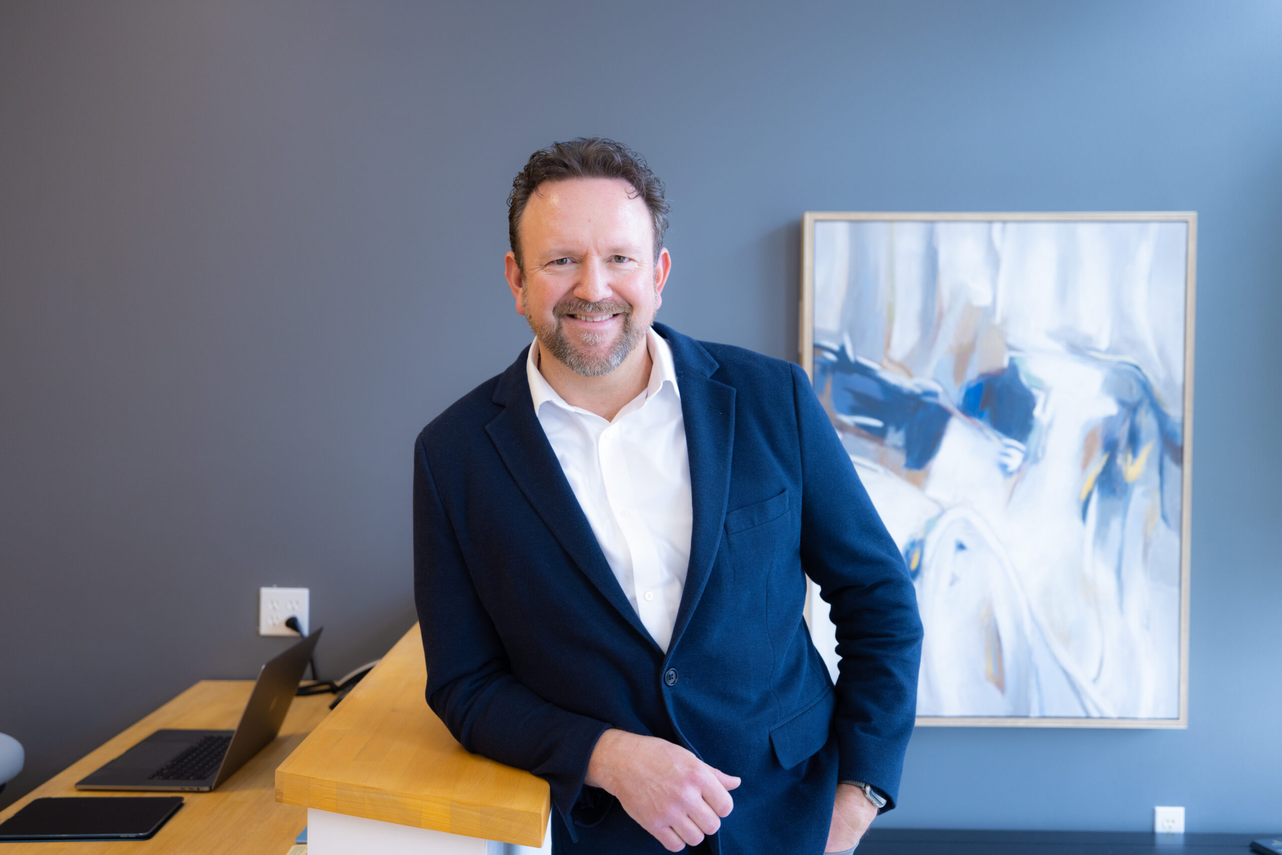
Your Doctor
Dr. Ryan Todd Lloyd
Doctor of Chiropractic and Qualified Medical Evaluator specializing in personal injury rehabilitation. With over 1,000 patients treated and a practice built entirely on lien-based care, Dr. Lloyd brings both clinical expertise and medicolegal understanding to every case.
His approach combines precise diagnosis — including cranial nerve and neurological examinations that most providers skip — with progressive, movement-based rehabilitation designed to achieve maximum medical improvement.
Ready to start your recovery?
Book your initial evaluation or call us directly. Same-week appointments available.