Introduction: The Patient Who Looks Fine on Film
I've sat across from patients who hand me their imaging reports with a mix of relief and frustration. Relief, because nothing looks broken. Frustration, because they're still in pain — and no one seems to believe them.
A radiologist writes "no acute osseous abnormality detected" and, for many patients, that sentence becomes a verdict. The insurance adjuster references it. The attorney references it. Sometimes, their own family references it. The message, spoken or not, is clear: if it doesn't show up on imaging, it probably isn't real.
This is one of the most damaging misconceptions in the evaluation and management of musculoskeletal trauma — particularly in low-velocity motor vehicle collisions. And it is contradicted by decades of peer-reviewed science.
The tissues most frequently injured (cervical ligaments, facet joint capsules, intervertebral discs, and neural structures) are soft tissues. They require specialized imaging, clinical assessment, or both to be identified. When we rely solely on plain films to validate or invalidate pain, we are using the wrong tool for the job — and patients pay the price.
Whiplash, more than any other mechanism, exposes this gap. It has been studied intensively, argued bitterly, and misrepresented frequently. What the literature actually shows us is clinically profound: pain after whiplash is real, measurable, and mechanistically explainable — even when X-rays are completely normal.
Part I: What Imaging Can and Cannot See

The Anatomy of a Low-Velocity Collision Injury
To understand why imaging misses what it misses, you have to start with what actually gets injured in a rear-end or low-speed collision.
In a typical rear-impact crash — even at speeds as low as 5–10 mph — the cervical spine undergoes a complex, non-physiological motion sequence. During the initial milliseconds of impact, the lower cervical segments extend while the upper segments flex. This creates an S-shaped deformation of the spine that does not occur in any natural movement.
The structures loaded during this motion include:
- Anterior longitudinal ligament — stretched or partially torn during hyperextension
- Posterior longitudinal ligament — compressed and impinged
- Facet joint capsules — stretched beyond physiological range, with documented capsular strains occurring at sub-fracture load levels
- Intervertebral discs — subjected to abnormal shear and compressive forces
- Cervical nerve roots and dorsal root ganglia — vulnerable to stretch injury, particularly at C5–C6 and C6–C7
- Suboccipital musculature and myofascial tissue — sustaining micro-tears and protective spasm
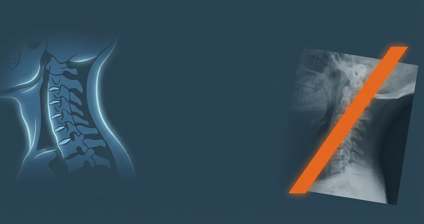
Of these, only gross bony disruption — fracture, dislocation, or advanced degenerative change — is reliably visualized on plain radiographs. Everything else is effectively invisible.
What Plain Radiographs Actually Show
Standard cervical spine X-rays are optimized for one thing: cortical bone. They show the density of mineralized tissue well and detect structural misalignment when it is large enough to be apparent on static film. They do not show:
- Ligament integrity
- Facet joint capsule health
- Intervertebral disc hydration or internal disruption
- Nerve root compression from soft tissue (as opposed to osteophyte)
- Muscle integrity
- Joint effusion at small synovial joints
CT scanning improves bone visualization significantly — particularly for detecting fractures missed on plain film — but it adds little to soft tissue assessment. A CT scan of a cervical spine with purely ligamentous or facet capsular injury will, in most cases, appear entirely normal.
What MRI Shows (and Still Misses)
Magnetic resonance imaging is a substantial improvement for soft tissue evaluation. MRI can detect disc herniation, disc signal change, ligamentous edema and partial tearing, and cord signal abnormality. In higher-grade whiplash injuries (WAD III and IV), MRI findings are more frequently positive.
However, standard clinical MRI protocols are not sensitive enough to detect all injury types relevant to whiplash.
Furthermore, the timing of MRI matters enormously. Soft tissue edema and signal change may resolve on MRI before symptoms resolve clinically — meaning a scan obtained weeks after injury can appear normal even when injury was present and pain persists. The imaging normalizes before the patient does.
Part II: The Biomechanics of Pain Without Imaging Findings

Facet Joint Injury — The Most Common Undetected Source
Of all the potential pain generators in whiplash, the cervical facet joint has the strongest evidence base. Research by Barnsley, Lord, and Bogduk — published across multiple peer-reviewed studies in the 1990s through 2000s — established that the cervical zygapophyseal (facet) joints are the single most common source of chronic pain after whiplash.
The facet joint capsule is richly innervated with nociceptors, and capsular strain at loads well below fracture threshold has been documented in cadaveric and in vitro models.
A critical paper by Panjabi et al. (1998) demonstrated that facet capsule strains exceed known physiological limits during simulated low-velocity rear impacts — even when no structural damage is visible. The nociceptors within the capsule are activated by strain, not structural failure. You do not need a torn capsule to produce pain; you need capsular deformation beyond its mechanoreceptor threshold.
Ligament Sprains and the Creep Phenomenon
Ligamentous tissue responds to sudden tensile loading in ways that are not captured on static imaging. The concept of creep — the progressive elongation of viscoelastic tissue under sustained load — is particularly relevant. When a ligament is loaded rapidly, as in a collision, it may undergo microfailure at the collagen fibril level without macroscopic rupture. This produces:
- Local inflammatory response
- Altered mechanoreceptor signaling
- Reduced joint position sense (proprioceptive deficit)
- Instability under dynamic loading
This is the crux of the clinical problem. The injury exists at a scale and in a tissue type that current standard-of-care imaging cannot resolve.
Neural Tension and Dorsal Root Ganglion Injury
The cervical nerve roots and dorsal root ganglia (DRG) are uniquely vulnerable in whiplash for several reasons. The DRG sits in the intervertebral foramen — a space that narrows during extension of the cervical spine. During the S-curve deformation of a rear impact, the foramen is dynamically compressed, and the DRG is subjected to compressive and tensile loads simultaneously.
Research by Ebraheim et al. and Winkelstein et al. (2001) demonstrated that DRG compression at sub-fracture loads produces sustained firing of nociceptive C-fibers — pain signaling — even after the compressive load is removed. This is a neurophysiological injury: the sensitization persists beyond the mechanical event.
Clinically, this presents as radiating pain, numbness, or paresthesia in a dermatomal distribution — findings that suggest nerve root involvement — in the presence of completely normal imaging.
Part III: Central Sensitization — When the Nervous System Becomes the Injury

Perhaps the most important concept in understanding persistent pain after low-velocity collision is central sensitization. It explains why some patients continue to experience significant pain long after tissue healing should have occurred — and why that pain is not fabricated, amplified, or psychological in origin. It is neurologically real.
The Science of Central Sensitization
Central sensitization refers to the amplification of neural signaling within the central nervous system — specifically, increased excitability of spinal cord neurons and changes in descending pain modulatory pathways. Key characteristics include:
- Allodynia: pain from stimuli that would not normally be painful (e.g., light touch)
- Hyperalgesia: exaggerated pain response to normally painful stimuli
- Referred pain expansion: pain spreading beyond the original injury site
- Temporal summation: repeated stimuli producing escalating pain responses
The Role of the Sympathetic Nervous System
Whiplash also affects the sympathetic nervous system in ways that contribute to pain and dysfunction. Research has shown altered sympathetic reactivity in chronic whiplash patients — including elevated resting sympathetic tone, reduced heart rate variability, and altered sudomotor response.
These autonomic changes are invisible on imaging. They require specialized testing to detect and are rarely captured in standard clinical workup — meaning patients presenting with symptoms like dizziness, visual disturbance, tinnitus, cognitive changes, and fatigue alongside neck pain may be dismissed as somatizing when they are in fact experiencing documented neurophysiological sequelae of their injury.
Part IV: The Epidemiology — How Common Is Pain Without Positive Imaging?
The Vehicle Damage Fallacy
One of the most persistent and damaging misconceptions in personal injury medicine is the assumption that low vehicle damage correlates with low occupant injury. The biomechanical reality is more complex.
Stiffer, more damage-resistant vehicles actually transfer more energy to occupants in low-speed impacts, because less energy is absorbed by vehicle deformation. A vehicle with a reinforced bumper designed to withstand a 5 mph impact without damage may transmit greater acceleration forces to the occupant than a vehicle whose bumper crumples — because the crumple zone dissipates energy that would otherwise reach the cabin.
Symptom Prevalence and Chronicity
Their chronicity is not explained by visible structural damage. It is explained by the mechanisms described above: facet sensitization, central sensitization, autonomic dysfunction, and proprioceptive deficit.
Part V: The Clinical Consequence — Patients Who Fall Through the Cracks
The Diagnostic Vacuum
When a patient presents with neck pain, headache, cognitive symptoms, and radiating upper extremity discomfort after a low-velocity collision — and their imaging is normal — they enter what I call a diagnostic vacuum. The objective findings that the healthcare system is conditioned to rely upon are absent. The subjective complaint is present. And increasingly, the burden shifts to the patient to prove their pain.
This has real consequences:
- Patients are undertreated or prematurely discharged from care
- Claims are denied or reduced based on imaging findings alone
- Patients are labeled as malingerers, catastrophizers, or secondary gain-seekers — often without any evidence to support those characterizations
- Psychological distress resulting from under-recognition compounds the neurological sensitization already present
The Clinical Examination: The Tool We're Not Using Enough
The answer to the imaging gap is not simply to order more imaging. It is to perform and document thorough clinical examination that captures what imaging cannot.
For the whiplash or low-velocity collision patient, a complete clinical assessment should include:
- Cervical range of motion — measured objectively with inclinometry
- Segmental motion palpation — assessing joint play and end-feel at each cervical level
- Neurological screen — dermatomal sensation, myotomal strength, deep tendon reflexes
- Pressure pain threshold testing — using algometry to quantify hyperalgesia
- Proprioceptive and balance assessment — particularly joint position error testing
- Upper limb tension testing — assessing neural tension and mechanosensitivity of the brachial plexus
- Cognitive and vestibular screening — particularly for patients reporting dizziness, cognitive fog, or visual disturbance
These assessments capture the functional injury that imaging cannot see. They provide objective data to support clinical findings. And they create a documentation record that reflects the actual clinical picture rather than the absence of a radiological one.
Part VI: Medicolegal Implications and the Standard of Care
Why This Matters Beyond the Clinical Setting
In personal injury practice, the relationship between imaging and legal outcome is significant — and frequently distorted. Defense experts and insurance companies routinely use negative imaging as the cornerstone of arguments that injuries are minor or non-existent. The strategy is effective precisely because it exploits a gap in public and professional understanding of what imaging can and cannot detect.
Clinicians practicing in the personal injury space must be prepared to articulate — clearly, in documentation, in reports, and if necessary in deposition — why a patient with normal imaging may have significant functional impairment. The science supports this position comprehensively. The failure is not in the patient's injury; it is in the diagnostic tool being misapplied as a final arbiter of injury presence.
The Physician's Ethical Obligation
There is an ethical dimension here that deserves direct statement. When we accept imaging as the final word on injury, we participate in the marginalization of patients whose injury is real but structurally invisible.
Part VII: Diagnostic Technologies That Close the Gap
While standard imaging has significant limitations, the landscape of diagnostic technology is evolving. Several tools show promise in detecting soft tissue injury that standard imaging misses:
High-Resolution MRI and Ligament Protocols
High-resolution MRI with specific ligament protocols — particularly for alar and transverse ligaments — demonstrates significantly improved sensitivity for upper cervical ligamentous injury. These protocols require longer scan times and specific expertise in interpretation, limiting their current clinical availability, but they represent an important advance.
Digital Motion X-Ray (DMX)
Digital Motion X-ray captures dynamic cervical motion — flexion, extension, lateral bending — in real time, allowing visualization of segmental instability and abnormal coupling patterns that static films cannot detect. While DMX is not universally accepted as a standard of care and interpretation requires expertise, it is increasingly used in the medicolegal context to document functional instability.
Quantitative Sensory Testing (QST)
QST provides objective measurement of somatosensory function — including pressure pain thresholds, thermal thresholds, and vibration detection. In research settings, QST has been used to objectively document and quantify central sensitization. It offers a reproducible, objective measure of neurological sensitization that cannot be dismissed as subjective.
Surface Electromyography (sEMG) and Functional Assessment Tools
Surface EMG measures muscle activation patterns and can detect asymmetries, altered firing sequences, and protective spasm patterns that indicate underlying joint or neural dysfunction. Paired with inclinometry and objective range of motion measurement, functional assessment provides a comprehensive, documentable picture of physical impairment.
Conclusion: A Different Lens
I opened with the patient who hands me their imaging report looking for answers. I want to close with what I tell that patient.
I tell them: the X-ray tells us what your bones look like. It tells us nothing about your ligaments, your facet joints, your nerve roots, or the way your nervous system has responded to this injury. Normal imaging is good news — it means nothing is broken. It does not mean nothing is wrong.
What we use to understand your injury is clinical examination — how your neck moves, where it restricts, what provokes your symptoms, how your nervous system is functioning. Those findings are real, they are documentable, and they guide your care.
The science is clear on this. Whiplash has been studied more rigorously than almost any other musculoskeletal injury mechanism. The research shows us, repeatedly and conclusively, that pain after low-velocity collision is mechanistically explainable, that the tissues responsible for that pain are largely invisible to standard imaging, and that absence of findings on radiograph or CT is not evidence of absence of injury.
Key References
- Barnsley L, Lord SM, Bogduk N. (1994). Comparative local anaesthetic blocks in the diagnosis of cervical zygapophyseal joint pain. Pain, 55(1):99–106.
- Castro WH, et al. (1997). No stress — no whiplash? International Journal of Legal Medicine, 109(5):236–240.
- Curatolo M, et al. (2001). Central hypersensitivity in chronic pain after whiplash injury. Clinical Journal of Pain, 17(4):306–315.
- Ivancic PC, et al. (2007). Whiplash causes increased laxity of cervical capsular ligament. Clinical Biomechanics, 22(10):1028–1034.
- Kaale BR, et al. (2005). Whiplash-associated disorders: A prospective study. Spine, 30(9):1073–1079.
- Lord SM, Barnsley L, Wallis BJ, Bogduk N. (1996). Chronic cervical zygapophysial joint pain after whiplash. Spine, 21(15):1737–1745.
- Panjabi MM, et al. (1998). Capsular ligament stretches during in vitro whiplash. Journal of Spinal Disorders, 11(3):227–232.
- Pfirrmann CW, et al. (2001). Magnetic resonance classification of lumbar intervertebral disc degeneration. Spine, 26(17):1873–1878.
- Radanov BP, et al. (1995). Two-year follow-up of patients with common whiplash injury. Journal of Neurology, Neurosurgery & Psychiatry, 59:565–568.
- Spitzer WO, et al. (1995). Scientific monograph of the Quebec Task Force on Whiplash-Associated Disorders. Spine, 20(8S):1S–73S.
- Sterling M, et al. (2003). Development of motor system dysfunction following whiplash injury. Pain, 103(1–2):65–73.
- Winkelstein BA, et al. (2001). The cervical facet capsule and its role in whiplash injury. Spine, 26(13):1508–1515.