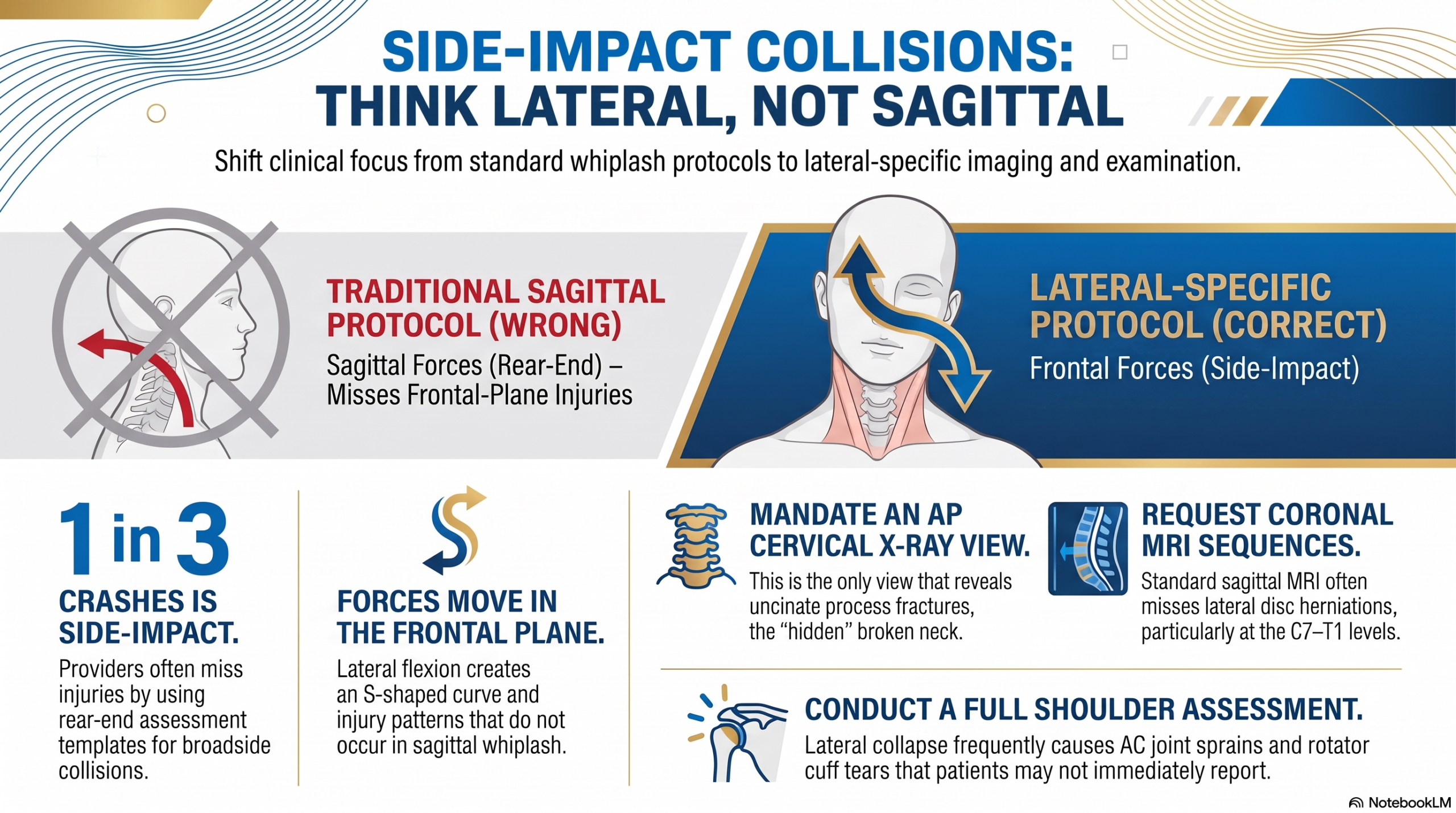
Side-impact crashes produce distinct, mechanism-specific injuries that rear-end collision protocols will not find.
Thirty-Four Percent
Thirty-four percent. More than one in three motor vehicle collisions in the United States is a side-impact crash. That number is not a footnote—it is a clinical mandate. Yet when providers walk into an exam room after a broadside collision, they frequently reach for the same assessment template they use for rear-end crashes: range of motion in the sagittal plane, tenderness along the posterior cervical spine, a standard lateral cervical X-ray series. They are looking for the right injuries in the wrong places.
Broadside collisions—properly called side-impact collisions, not “T-bones”—do not produce the same injury patterns as rear-end crashes. The forces travel in a different plane. The body collapses in a different direction. The structures at risk are different. And the imaging needed to find those structures requires deliberate protocol changes that most providers never make.
The result is a pattern of missed injuries, delayed diagnoses, and patients who are told their pain is disproportionate to the accident—when in fact no one has ordered the imaging that would find what’s wrong. This article explains the mechanism, identifies the injuries, and outlines the clinical and imaging protocols that close the gap.
“They are looking for the right injuries in the wrong places.”
The Mechanism: Forces in the Frontal Plane
To understand why broadside collisions create distinct injury patterns, you have to understand the difference between sagittal-plane and frontal-plane loading of the cervical spine.
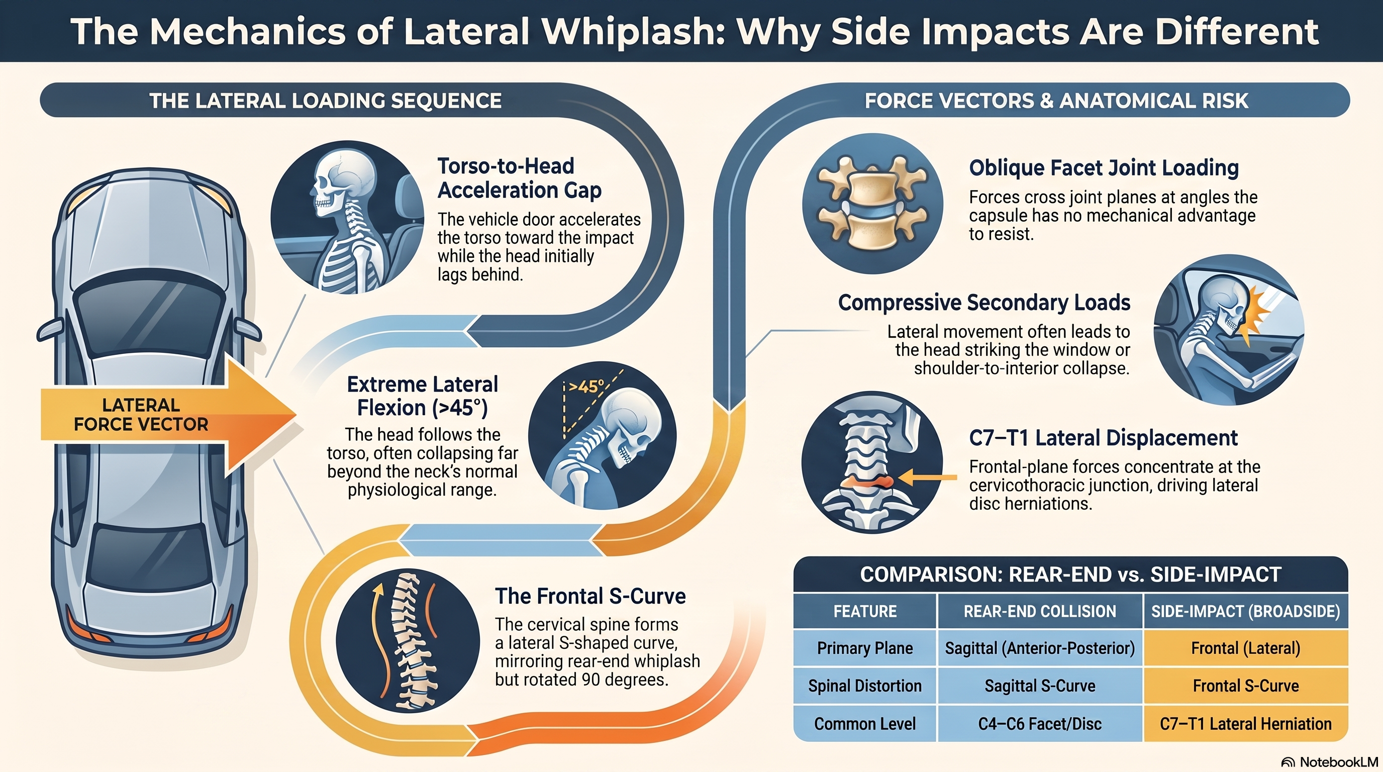
In a rear-end collision, forces travel along the anterior-posterior axis. The head first lags behind the accelerating torso (extension), then snaps forward (flexion). This is the classic whiplash sequence, well documented in biomechanical literature since the Grauer 1997 Spine study and reinforced by the 2016 Elliott Physical Therapy Journal research on the S-shaped cervical curve. Providers trained in rear-end mechanics know to look for posterior disc herniations, facet capsule injuries at the C4–C6 levels, and the S-shaped curve distortion on dynamic imaging.
In a broadside collision, the force vector is lateral. The body moves toward the impacted side while the head initially lags—then follows, often far beyond physiological range in the frontal plane. The result is:
- Lateral flexion toward the impact side, frequently exceeding the neck’s normal range of 45 degrees
- An S-shaped cervical curve in the frontal plane (not the sagittal plane), as documented in the same biomechanical literature applied to whiplash
- Potential for the head to strike the side window, adding a compressive load on top of the lateral flexion
- Shoulder loading when the passenger collapses laterally onto the shoulder complex
- Forces that cross joint planes obliquely—creating injury patterns that don’t occur in rear-end mechanics
Key distinction: This is not a minor variation of whiplash. It is a fundamentally different loading event in a different anatomical plane.
Lateral Disc Herniations: The Injury Your MRI Protocol May Be Missing
In rear-end collisions, disc herniations are predominantly posterior or posterolateral. They appear on standard sagittal MRI sequences and are relatively straightforward to identify. In broadside collisions, the frontal-plane forces drive herniations in the lateral direction—most commonly at the C7–T1 levels, where lateral flexion forces concentrate.
Here is the clinical problem: standard MRI protocols for the cervical spine are built around sagittal sequences. They are optimized to see what happens in the anterior-posterior axis. Lateral herniations at C7–T1 can be subtle on sagittal images, partially hidden by the shoulder girdle on standard sequences, or missed entirely when no one has told the radiologist to look for lateral pathology.
What changes clinical outcomes in these cases:
- Request coronal MRI sequences explicitly. A radiologist reviewing a standard cervical MRI without coronal cuts may report a normal study while a lateral herniation at C7–T1 is present but not visualized.
- Communicate the mechanism to the radiologist. “Side-impact collision with lateral flexion mechanism” changes what the radiologist is looking for. Without that communication, they apply a standard protocol to a non-standard injury.
- Correlate the imaging with the clinical presentation. Upper extremity symptoms—pain, numbness, or weakness tracking into the arm on the impact side—in a broadside collision patient should be treated as a lateral herniation until proven otherwise, even if the initial MRI read is “no significant pathology.”
The lesson from rear-end collision research applies here: a normal MRI report is not the same as a normal MRI. It means the imaging protocol used did not find pathology. That is not the same thing.
Uncinate Process Fractures: The Hidden Broken Neck
This is the finding that most providers miss entirely, and the one that has the most significant clinical and medicolegal implications.
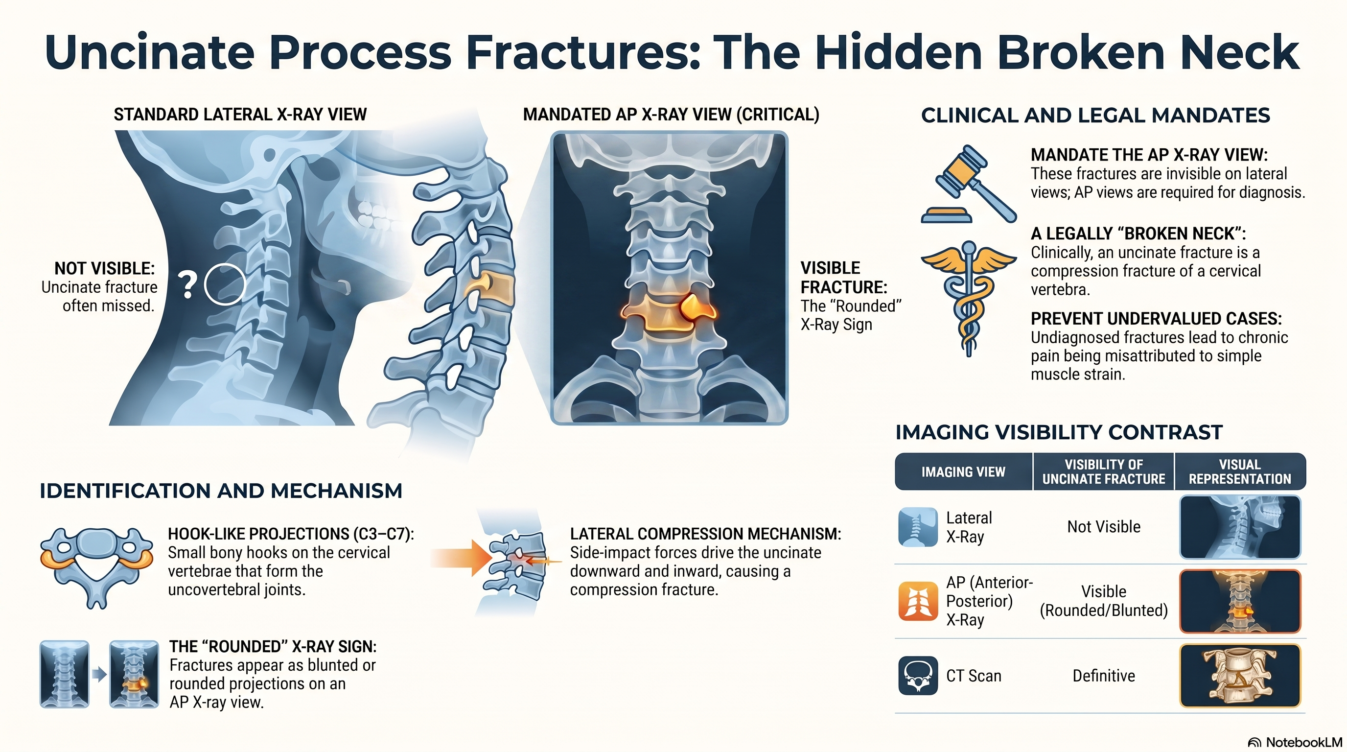
The uncinate processes are small hook-like projections on the superior lateral margins of the cervical vertebral bodies, from C3 through C7. They form the uncovertebral joints (joints of Luschka) and play a role in guiding cervical motion and protecting the intervertebral foramina. In normal anatomy, they appear as sharp, well-defined bony points on anterior-posterior (AP) cervical X-ray views.
In a side-impact collision, the lateral compressive forces that accompany lateral flexion can fracture the uncinate process. The fracture pattern is a compression fracture—the uncinate is driven downward and inward by the collapsing vertebral bodies. On imaging, the finding is subtle:
- On AP cervical X-ray: loss of the normal sharp point of the uncinate process; instead, the projection appears rounded or blunted. The space at the uncovertebral joint may appear asymmetrically narrowed.
- On lateral cervical X-ray: the fracture is not visible. This is the critical point. Standard cervical X-ray series often consist of lateral and oblique views. The AP view—the only view that reveals uncinate fractures—is frequently omitted or deprioritized.
- On CT: the fracture is definitive. If clinical suspicion is high and X-ray findings are equivocal, CT is the appropriate next step.
Medicolegal note: An uncinate process fracture is a compression fracture of a cervical vertebra. It qualifies, legally, as a broken neck. Patients with uncinate fractures who are never diagnosed have cases that are undervalued from the first day—because no one ordered the AP view.
The clinical mandate is simple: For every broadside collision patient, the cervical X-ray series must include an AP view. Not as an optional add-on—as a required component of the protocol. Compare the right and left uncovertebral joints on that AP view. Look for asymmetric narrowing. Look for rounded projections where sharp points should be. When in doubt, order CT.
“The AP view is the only one that reveals uncinate fractures. It is the one most providers skip.”
Facet Joint Injuries: When the Force Vector Crosses the Joint Plane
Cervical facet joints are positioned to bear load and guide motion primarily in the sagittal plane. Research has established that facet joints account for approximately 55% of chronic neck pain following whiplash injuries (Lord et al., published in Spine). Those findings come from rear-end collision populations.
In a side-impact collision, the force vector crosses the facet joint planes at angles that don’t occur in sagittal-plane loading. The oblique orientation of cervical facets means that frontal-plane forces create capsular shear loads that exceed those seen in rear-end mechanics. The capsule—already the most innervated structure in the facet joint complex—is loaded in a direction it is not mechanically designed to resist.
The clinical implication: facet-mediated pain in a broadside collision patient may be more severe and more refractory than in a comparable rear-end collision patient, because the injury mechanism is less physiological. The same diagnostic approaches apply—medial branch block patterns, characteristic referred pain maps—but the provider should not be reassured by the absence of sagittal-plane motion restriction. Lateral flexion restriction, and pain with combined lateral flexion and rotation, are more sensitive indicators in side-impact facet injury.
Shoulder Complex Trauma: The Injury No One Examines
When the body collapses laterally in a broadside collision, the shoulder on the impact side bears load in a way that the shoulder was not designed to handle. Patients may not spontaneously report shoulder pain in the acute phase—they are overwhelmed by neck pain, headache, and the neurological symptoms of their cervical injury. But the shoulder examination is not optional.
Injury patterns to evaluate and document:
- Acromioclavicular joint sprain: The AC joint is vulnerable to superior shear loads, which is exactly what occurs when the shoulder is driven downward into a lateral collapse. Tenderness directly over the AC joint, pain with cross-body adduction, and the piano key sign are the key examination findings.
- Rotator cuff injury: Compressive loading of the shoulder during lateral collapse can create partial or full-thickness rotator cuff tears. The supraspinatus is most vulnerable. In patients over 40, a negative initial exam does not rule out rotator cuff pathology; MRI of the shoulder may be necessary.
- Glenohumeral joint trauma: Posterior capsule injury and posterior labral tears can result from the compressive lateral loading. These are frequently missed because they do not cause the classic anterior instability signs.
- Clavicle fractures: Less common but possible when compressive forces are concentrated through the shoulder girdle.
Documentation imperative: The causation argument connecting the lateral collapse mechanism to a specific shoulder injury depends on the provider having examined and documented the shoulder from the first visit. Providers who examine only the cervical spine create a gap in the causation chain that becomes very difficult to close later.
The Pituitary Gland Difference: A Counterintuitive Finding
One of the more surprising distinctions between rear-end and broadside collisions involves the pituitary gland.
In a rear-end collision, the brain translates forcefully within the skull in the anterior-posterior direction. The pituitary gland—housed in the bony sella turcica at the base of the skull—can be driven against the posterior wall of that bony cup. The result is pituitary stalk injury, which can manifest as hormonal dysfunction: menstrual irregularities, fatigue, changes in libido, mood disturbance, and in severe cases, diabetes insipidus or growth hormone deficiency.
In a broadside collision, the brain’s primary motion is lateral—side to side within the skull. The pituitary gland, in lateral brain motion, does not impact the bony sella with the same force. It swings laterally within the sella rather than being driven against its posterior wall. The clinical takeaway: hormonal dysfunction following a motor vehicle collision is more characteristic of rear-end mechanisms than broadside mechanisms. This does not mean pituitary injury cannot occur in a side-impact crash—traumatic brain injury of any mechanism can affect the hypothalamic-pituitary axis—but when a provider is building a causation argument linking hormonal dysfunction to a specific collision mechanism, the directionality matters. Rear-end collision is the higher-risk mechanism for this specific sequela.
The Imaging Protocol That Changes Everything
The single highest-yield clinical change in broadside collision care is protocol-level: mandate an AP cervical X-ray view for every side-impact patient, and explicitly communicate the mechanism to the radiologist for every MRI ordered.
Cervical X-Ray Protocol for Broadside Collisions
- AP view: Required. This is the only view that visualizes uncinate fractures. Compare bilateral uncovertebral joints. Document any asymmetric narrowing or blunting of uncinate projections.
- Lateral view: Required. Evaluate lordosis, disc height, prevertebral soft tissue.
- Obliques: Evaluate foraminal stenosis, which may be relevant given the lateral disc herniation pattern.
- Open mouth (odontoid) view: Evaluate the atlantoaxial complex, particularly relevant if head-window contact occurred.
MRI Protocol for Broadside Collisions
- Request coronal sequences in addition to standard sagittal sequences. This is the view that reveals lateral herniations.
- Specify the mechanism on the order: “Side-impact MVA, lateral flexion mechanism, evaluate for lateral disc herniation at C7–T1 and uncinate pathology.”
- If C7–T1 is undervisualized due to shoulder artifact, request a cervicothoracic junction protocol or repeat imaging with adjusted shoulder positioning.
- Shoulder MRI: Order as a dedicated study when shoulder examination findings are present. Do not assume the shoulder MRI is optional because the cervical MRI is pending.
Digital measurement on the AP view, rather than visual estimation, improves sensitivity for subtle uncinate asymmetry. If your practice uses digital radiograph software, apply measurement tools to document the bilateral uncovertebral joint spaces numerically.
Clinical Examination: What to Look for in the Room
The physical examination in a broadside collision patient should be systematically expanded beyond the standard whiplash protocol.
Cervical Range of Motion
Lateral flexion is the critical measurement. In a rear-end collision patient, lateral flexion is often preserved while sagittal-plane motion is restricted. In a broadside collision patient, lateral flexion toward the impact side is frequently severely limited and painful. Document it bilaterally and note the asymmetry. Coupled motions—lateral flexion combined with rotation—often reveal restrictions that single-plane measurements miss.
Neurological Examination
Upper extremity neurological testing is essential. The lateral herniation pattern at C7–T1 affects the C8 and T1 nerve roots—producing symptoms in the medial forearm, ring finger, and small finger, as well as intrinsic hand weakness. Providers applying a standard cervical exam may test C5–C7 root function while missing the C8–T1 involvement that characterizes side-impact disc herniation levels.
Shoulder Examination
Conduct a full shoulder assessment regardless of patient complaint. Examine: AC joint palpation, impingement testing (Neer, Hawkins), rotator cuff strength testing (supraspinatus, infraspinatus, subscapularis), cross-body adduction for AC joint involvement, and glenohumeral range of motion.
Posture and Gait
Observe the patient standing. A lateral weight shift away from the impact side, shoulder elevation on the impact side (guarding), and antalgic gait if lower extremity involvement is present are mechanism-consistent findings that should be documented.
The Seatbelt Sign
Inspect for diagonal bruising across the chest from the shoulder belt. In a broadside collision, the belt restraint creates torsional forces across the thorax as the lateral collapse is arrested by the diagonal shoulder belt. Look for rib tenderness along the belt line and document any skin markings.
Documentation: Building the Causation Case
Understanding mechanism tells you what to look for. Clinical documentation translates that understanding into the legal and insurance record.
For every broadside collision patient, the mechanism description in your initial report should specify:
- Which side was impacted (driver side or passenger side)
- Approximate speed and angle of impact if known
- Whether the head struck the side window
- Seatbelt use and presence of seatbelt sign
- Time of onset (immediate vs. delayed)
- Vehicle deformation and deployment of side-curtain airbags, if documented
The vector analysis portion of your report should connect the frontal-plane force vector to the specific structures injured. “Frontal-plane lateral flexion forces at C7–T1 are consistent with lateral disc herniation at that level” is a causation statement that holds up. “Patient was in a motor vehicle accident and has neck pain” does not.
When documenting the shoulder, tie the injury explicitly to the lateral collapse mechanism: “Lateral collapse of the torso toward the impact side, combined with seatbelt restraint, is consistent with compressive loading of the right acromioclavicular joint, producing the grade II AC sprain documented on examination.”
Why These Injuries Are Consistently Missed
The missed injury rate in broadside collision cases is not random. It follows a predictable pattern driven by four systemic failures:
- Training bias: The majority of whiplash biomechanics research—and therefore the majority of clinical training—focuses on rear-end mechanisms. Providers pattern-match to what they learned, and what they learned was sagittal-plane mechanics.
- Terminology confusion: “T-bone” is a colloquial term that communicates nothing about mechanism. The clinically precise term—“side-impact collision with frontal-plane lateral flexion mechanism”—is what belongs in your documentation and on your imaging orders. Language shapes what gets looked for.
- Imaging protocol defaults: Radiology departments use default protocols. Unless the ordering provider specifies the mechanism and requests non-standard sequences, the radiologist applies a standard cervical MRI protocol. Standard protocols are not designed for lateral herniation visualization. The responsibility for protocol modification falls on the ordering provider, not the radiologist.
- Patient presentation patterns: Patients in acute pain from a broadside collision present primarily with cervical and head complaints. A provider who waits for the patient to spontaneously report shoulder or hand symptoms will miss them. The examination must actively look for them.
Conclusion: Thirty-Four Percent of Your Patients Deserve a Different Protocol
One in three motor vehicle collision patients in your practice was in a side-impact crash. They did not have a rear-end collision. They did not have a sagittal-plane loading event. They have a different set of vulnerable structures, a different imaging protocol requirement, and a different examination emphasis than the rear-end collision patient in the next room.
The injuries—lateral disc herniations at C7–T1, uncinate process fractures on the AP view, facet capsule injuries from oblique force vectors, shoulder complex trauma from lateral collapse—are not rare or exotic. They are mechanism-predictable. When you understand the force vector, you know what to look for. When you know what to look for, you find it. When you find it, you document it. When you document it, the causation chain is intact.
Thirty-four percent of your motor vehicle collision patients have these mechanisms. The protocol changes are not complicated: add the AP view, request coronal MRI sequences, communicate the mechanism to radiology, examine the shoulder, and describe the frontal-plane force vector in your reports. These are not extraordinary measures. They are what the mechanism demands.
“Thirty-four percent of your patients deserve a different protocol. Most don’t get it because providers apply rear-end templates to side-impact bodies.”
References
- Grauer JN, Nightingale RW, Pintar FA, et al. Whiplash produces an S-shaped curvature of the neck with hyperextension at lower levels. Spine. 1997;22(21):2489–2494.
- Elliott JM, et al. Characterization of acute and chronic whiplash-associated disorders. Journal of Orthopaedic & Sports Physical Therapy. 2016. (Verify full volume and page numbers before publication.)
- Lord SM, Barnsley L, Wallis BJ, Bogduk N. Chronic cervical zygapophysial joint pain after whiplash. Spine. 1996;21(15):1737–1745.