Bulge vs. Herniation: What Your MRI Report Really Means — and Why the Difference Can Change Everything
One word on a radiology report can be worth tens of thousands of dollars. Not because of the test, and not because of the injury itself — but because of whether the finding on that report meets a specific definition that determines whether permanent impairment can be established, whether the injury qualifies for an impairment rating under the AMA Guides, and whether the legal value of a post-collision case is calculated in the thousands or the hundreds of thousands.
That word is “herniation.”
The problem is that most patients leave their imaging appointments with a piece of paper that uses terms like “disc protrusion,” “disc bulge,” “broad-based herniation,” and “focal protrusion” without any explanation of what those terms mean, how they differ from each other, or what their clinical and legal implications are. And in many cases, the terminology on the report itself is imprecise — using “herniation” to describe what is technically a bulge, or “protrusion” without specifying whether it meets the criteria for pathological herniation.
This is not a trivial inconsistency. The distinction between a disc bulge and a disc herniation — defined precisely by one of the most widely cited research standards in spinal radiology — separates degenerative wear from traumatic injury, eliminates the possibility of an impairment rating from the possibility of establishing permanent impairment, and fundamentally changes the medical, clinical, and legal narrative of a post-collision case. Every provider who treats post-collision patients and every patient who has ever been told their spine shows “just some degeneration” needs to understand the difference.
The Gold Standard: What the Fardon Paper Actually Says
The most widely cited framework for lumbar disc nomenclature is the classification system established by Fardon and colleagues, most recently updated in collaboration with the American Society of Neuroradiology, the American Society of Spine Radiology, and the North American Spine Society. The Fardon paper is the standard against which all disc terminology should be measured — and it provides precise, measurable definitions based on a single anatomical criterion: the circumferential extent of disc displacement.
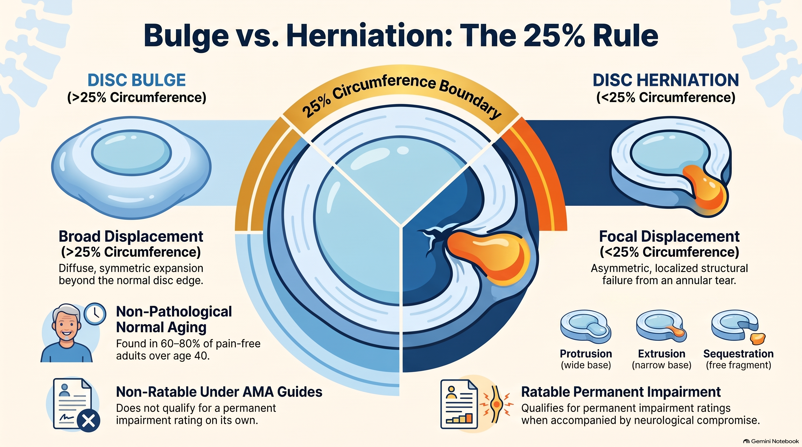
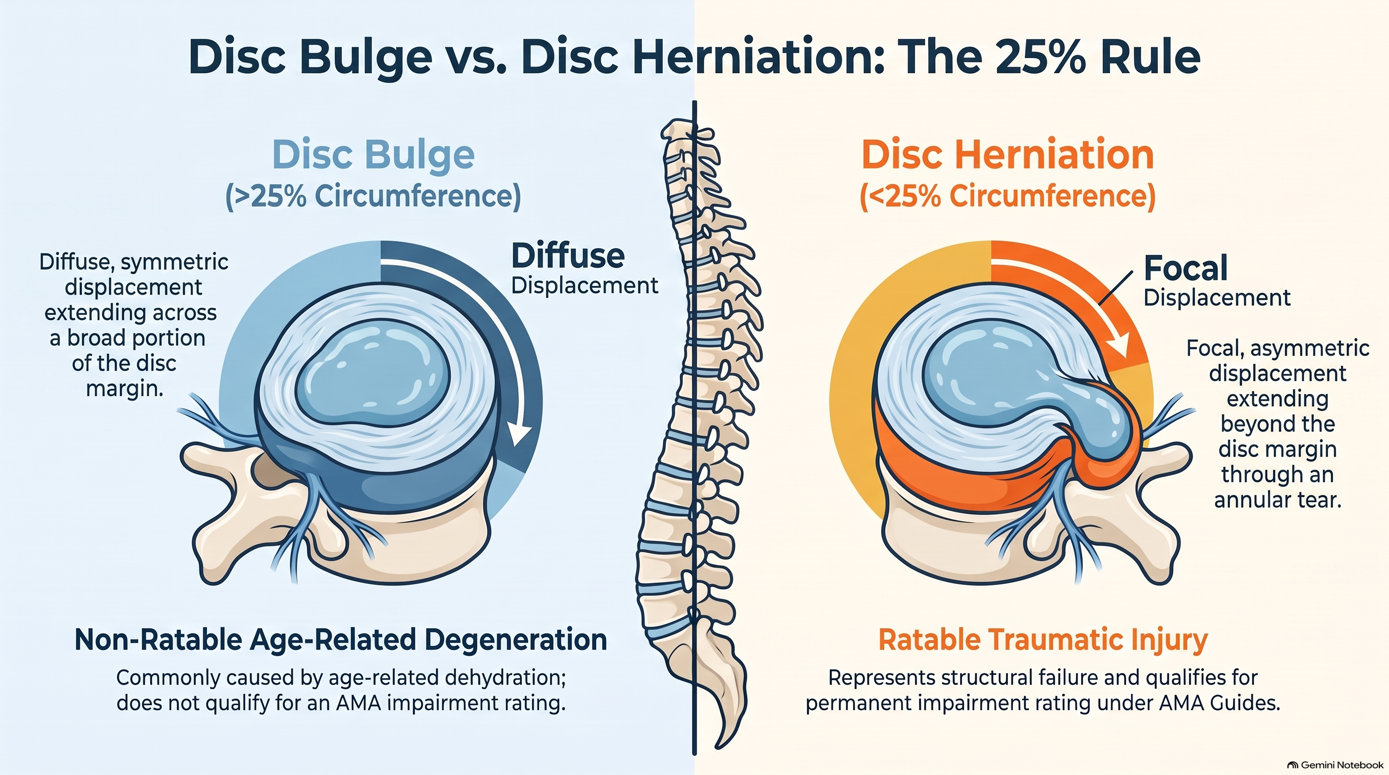
The definition is straightforward. A disc bulge involves displacement of disc material beyond the normal disc margins encompassing more than 25% of the disc circumference. A disc herniation involves focal displacement of disc material beyond the normal disc margins encompassing less than 25% of the disc circumference.
This is a counterintuitive distinction for many patients. A bulge — which sounds more diffuse and less serious — actually involves a larger portion of the disc margin. A herniation — which sounds more dramatic — is defined by its focal, asymmetric nature, involving a smaller and more concentrated zone of displacement.
The clinical significance of this distinction flows directly from its geometry. A bulge is, by definition, a symmetric or near-symmetric finding. It is common, often related to age-related disc dehydration and loss of disc height, and in most cases does not qualify as pathological in the medico-legal sense. A herniation, by contrast, is focal. It is asymmetric. It is more likely to represent a specific event — a tear in the annular fibers, a displacement of nuclear material through that tear — and it is the finding that qualifies for impairment rating under the American Medical Association Guides to the Evaluation of Permanent Impairment.
“One number separates normal aging from ratable permanent injury: 25% of the disc circumference.”
Bulge: What It Is and What It Isn’t
A disc bulge is among the most common findings on spinal MRI, and among the most frequently misunderstood. Studies examining asymptomatic volunteers — individuals with no back pain, no neck pain, and no history of injury — consistently find bulges in a majority of subjects by middle age. Research published in the American Journal of Neuroradiology documented degenerative findings including disc bulges in approximately 60 to 80 percent of asymptomatic individuals over the age of 40.
This prevalence is the clinical reason why a bulge, by itself, does not qualify for impairment rating under the AMA Guides. A finding present in the majority of the pain-free population cannot, by itself, establish the presence of injury, permanent impairment, or medical necessity for extended treatment. The AMA Guides require that a ratable finding represent a departure from normal — and a bulge, however symptomatic it may be, does not meet that threshold.
This does not mean that bulges are clinically insignificant. A bulge at a level corresponding to a patient’s reported pain pattern, following trauma with a mechanism that would produce axial loading or flexion-compression forces, is a clinically meaningful finding — especially when the adjacent levels are normal. If a finding is truly age-appropriate, it should be present at multiple levels, reflecting the systemic effect of aging on disc tissue. A single-level bulge at L4-5 with normal findings at L3-4 and L5-S1 is not age-appropriate. It is level-specific. And level-specific findings have level-specific causes.
The patient-facing explanation is useful: imagine pressing down on a hamburger patty. The meat spreads outward around the full circumference. The spreading is even, diffuse, and occurs around the entire margin. That is a bulge. It is common, it can cause pain, and it may require treatment — but under the current classification system, it does not meet the threshold for permanent impairment.
Herniation and Its Subtypes: Not All Herniations Are Equal
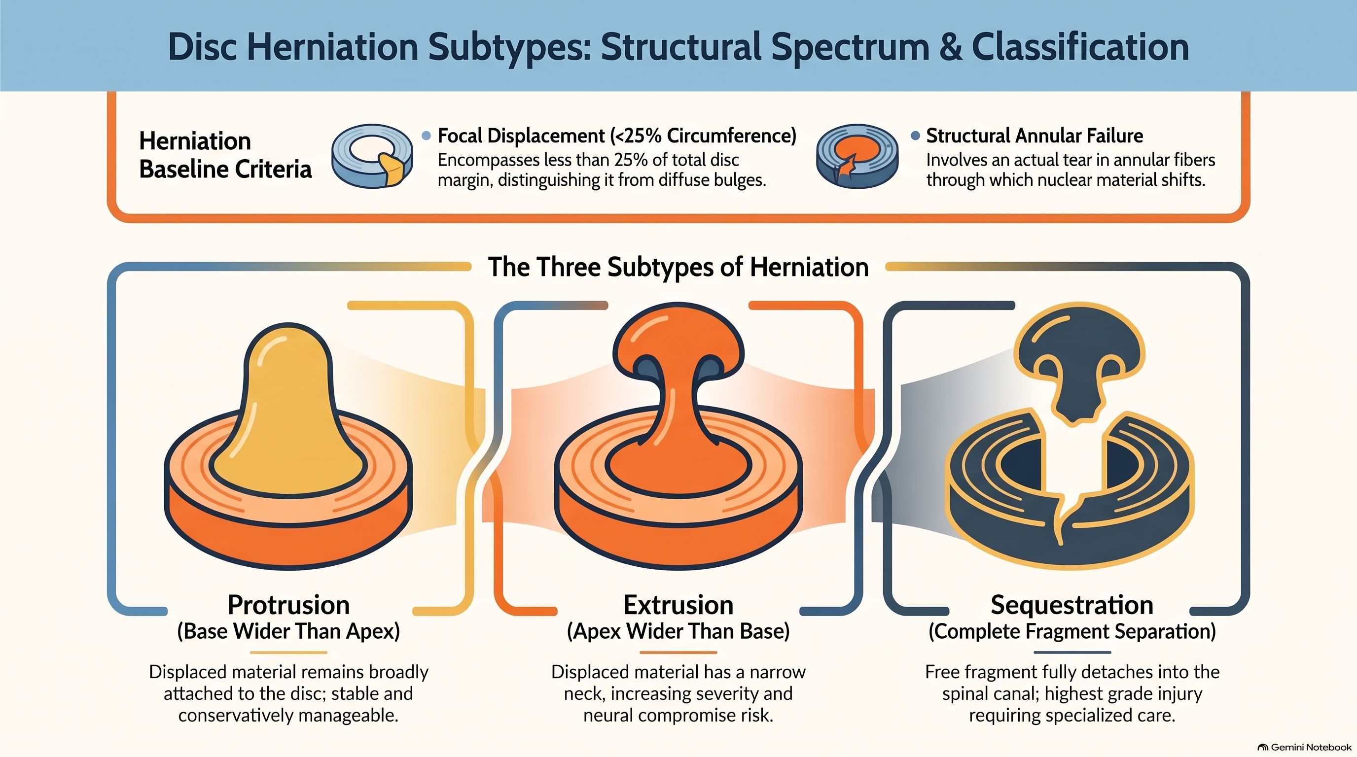
A herniation is a focal displacement — less than 25% of the disc circumference — in which disc material extends beyond the normal margin of the disc. Unlike a bulge, a herniation represents a structural failure: a tear in the annular fibers through which nuclear material is displaced. The nature of that displacement, and the relationship between the displaced material and its source, determines the subtype.
Protrusion is the first and most common herniation subtype. In a protrusion, disc material extends beyond the disc margin, but the base of the herniation — the portion still connected to the disc — is wider than the apex. Visualize a mushroom with a wide, thick stem: the material that has displaced is still broadly connected to the parent disc. Protrusions are relatively stable, less likely to sequestrate, and typically more amenable to conservative management. They are, nonetheless, herniations — they meet the circumferential criterion, and they qualify for impairment rating when they produce neural compromise or meet the size thresholds specified in the AMA Guides.
Extrusion represents a more advanced herniation subtype. In an extrusion, disc material has pushed through a defect in the annulus in such a way that the apex of the displaced material is wider than the base — the neck of the herniation is narrower than the head. Visualize a mushroom with a thin, narrow stem: the displaced material is only loosely attached to the parent disc and may be difficult to reduce through conservative means. Extrusions are generally more serious than protrusions, carry a higher risk of further migration, and are more likely to produce significant neural compromise.
Sequestration represents the most advanced herniation state. In sequestration, disc material has completely separated from the parent disc and exists as a free fragment within the spinal canal. This fragment cannot reabsorb through the normal mechanisms available to contained herniations. It cannot be reduced through conservative care. It may migrate up or down the canal, producing variable and sometimes fluctuating neurological symptoms. Sequestration is frequently cited as a surgical indication, and in the context of post-collision injury, it represents the highest grade of disc injury recognizable on imaging.
Internal Disc Disruption: The Injury the MRI Doesn’t Always See
One of the most important and least-discussed categories of disc injury is internal disc disruption — a condition in which the internal architecture of the disc is damaged without any material extending beyond the vertebral margins. In internal disc disruption, the annular fibers tear from within, typically in the posterior third of the annulus, where pain fiber density is highest. Nuclear material may track into these tears, producing a characteristic signal change on MRI — increased signal on T2-weighted sequences, sometimes visible as a “high intensity zone” — but the disc itself does not extend beyond its normal margins.
This matters for several reasons. First, internal disc disruption can produce significant and persistent axial pain without producing any finding that a standard radiology read would call a herniation. The disc looks, from the outside, essentially intact. The annular disruption is internal. A radiologist reading the study without specific attention to posterior annular signal may report “no herniation” and leave it at that. Second, the posterior third of the annulus has the highest concentration of nociceptive nerve fibers of any region of the disc. Tears in this region are not silent injuries — they are a well-documented source of severe, chronic discogenic pain that can be causally related to trauma without producing any imaging finding that triggers the herniation classification.
The clinical implication is that a normal-appearing disc on MRI is not the same as an uninjured disc. The finding of internal disc disruption requires attention to T2 signal characteristics, correlation with the clinical presentation of axial pain without radiculopathy, and in some cases provocative discography when the clinical picture warrants it.
Clinical note: A “high intensity zone” on T2-weighted MRI in the posterior annulus is a documented marker of internal disc disruption. It does not meet herniation criteria but is clinically significant and should be correlated with the patient’s pain pattern and collision mechanism.
The Age-Appropriate Problem: A Challenge Worth Making
The phrase “age-appropriate changes” appears on MRI reports with enough frequency that it has become nearly invisible — a dismissal embedded in the language of radiology that patients, attorneys, and even treating clinicians often accept without scrutiny. The challenge to this language is well-founded and important.
If a degenerative finding — a disc bulge, a reduction in disc height, a loss of T2 signal indicating desiccation — is truly age-appropriate, it should be present at multiple levels. The effects of aging on disc tissue are systemic, not level-specific. The intervertebral discs throughout the spine undergo the same biochemical aging processes: loss of proteoglycan content, reduction in water-binding capacity, gradual loss of disc height. A spine that is aging normally would show these changes at multiple levels, not one.
A single-level finding at L4-5, with normal findings at L3-4 and L5-S1, is not age-appropriate. It is level-specific. And level-specific findings have level-specific causes. When a patient was involved in a motor vehicle collision three months before the MRI was taken, and the level-specific finding correlates with the patient’s reported pain distribution, the radiology report’s characterization of that finding as “age-appropriate” is not a neutral statement — it is an incorrect one. Challenging that characterization, with the patient’s clinical presentation and the specificity of the imaging finding as the basis, is both appropriate and clinically necessary.
Direction of Herniation: Mechanism Matters
The direction in which disc material displaces is not random — it is related to the mechanism of the injury and the direction of forces applied to the disc at the moment of impact. In rear-end collisions, the hyperextension-hyperflexion mechanism produces posterior and posterolateral displacement of disc material. The rapid extension phase loads the posterior annulus, and if the annular fibers fail, the herniation will emerge posteriorly — centrally or paracentrally, at the midline or just to one side. This pattern aligns with the most common clinical presentation of post-collision disc injury: central or paracentral herniation producing cord impingement or nerve root compression at the affected level.
Broadside collisions produce a different pattern. The lateral forces applied in a side-impact collision produce lateral disc displacement at the cervical and upper thoracic levels, often requiring coronal MRI sequences to fully visualize. A lateral foraminal herniation following a left-side impact, documented on coronal sequences, is mechanistically consistent with the collision in a way that supports causal attribution.
This anatomical correlation — mechanism of collision, direction of force, direction of herniation — is part of the comprehensive documentation strategy that transforms a radiology finding from an incidental observation into a documented injury with an established cause.
The Impairment Rating and Legal Valuation
The clinical and legal consequences of the bulge-versus-herniation distinction are concrete and significant. Under the AMA Guides, a disc bulge — by itself — does not qualify as a ratable finding. A disc herniation, meeting the Fardon circumferential criteria and producing documented neurological compromise or meeting size thresholds, does qualify.
The practical effect is that a case with a documented disc herniation that meets impairment criteria can support a permanency claim — a finding of lasting structural change expected to cause ongoing impairment and future medical needs. A case documented only as “mild degenerative changes” and “disc bulge at L4-5” cannot. The same patient, the same collision, the same symptoms — the documented terminology determines whether permanent impairment can be established.
This is why the terminology on the radiology report matters, and why providers who treat post-collision patients must be prepared to review imaging directly rather than accepting radiology report language as the final word. A radiologist who uses “broad-based herniation” to describe a finding that covers more than 25% of the disc circumference has produced a term that is internally inconsistent by the Fardon standard: a broad-based finding is a bulge, not a herniation. A provider who reviews the images and documents the circumferential extent of the finding using Fardon nomenclature in the clinical record has produced documentation that is both more precise and more legally defensible than the original radiology report.
The Documentation Standard
When the radiology report is unambiguous: Echo the language, connect the finding to the mechanism of injury, and document neural involvement.
When the radiology report is ambiguous: Correct the classification, document the image review directly, and state the finding in Fardon-compliant terms. Every disc injury should include: level, classification, direction, neural compromise, clinical correlation, and causal relationship to the collision.
Precise clinical documentation in post-collision disc injury cases requires more than citing the radiology report. When the radiology report is unambiguous — clearly identifying a focal herniation with circumferential and dimensional measurements — the clinical note should echo that language, connect the finding to the mechanism of injury, and document the neural involvement. When the radiology report is ambiguous — using terms like “protrusion” or “broad-based herniation” without meeting the Fardon circumferential criterion — the clinical note should correct the classification, document the image review directly, and state the finding in Fardon-compliant terms.
Every disc injury documented in a post-collision clinical record should include: the level, the classification (bulge, herniation, protrusion, extrusion, or sequestration), the direction of displacement, the presence or absence of neural compromise, the correlation with the clinical examination and symptom pattern, and the causal relationship to the documented collision mechanism. This documentation protects the patient’s right to an accurate diagnosis, supports appropriate treatment authorization, and provides the evidentiary foundation for any impairment or legal determination that follows.
Conclusion
The distance between a disc bulge and a disc herniation is less than the width of a single anatomical margin — but the clinical, legal, and financial distance between those two findings is enormous. The Fardon classification provides a precise, measurable, research-supported standard for making that distinction. The 25% circumferential criterion is not a gray area. It is a line, and which side of that line a finding falls on determines whether a patient qualifies for an impairment rating, whether a permanency claim can be supported, and whether a post-collision case is valued in the thousands or the hundreds of thousands.
Most patients leave their imaging appointments without understanding any of this. They hear “there’s some disc degeneration” and accept it as a verdict — when what they should hear is a precise classification, a correlation with their mechanism of injury, and a clear explanation of what that finding means for their treatment and their future. The clinician who understands the Fardon standard, reviews imaging directly, uses correct nomenclature in the clinical record, and explains the distinction clearly to patients is not simply being more accurate. They are advocating for a patient who cannot advocate for themselves.
References
- Fardon DF, Williams AL, Dohring EJ, et al. Lumbar disc nomenclature: version 2.0. Spine. 2014;39(24):E1448–E1465.
- Brinjikji W, Luetmer PH, Comstock B, et al. Systematic literature review of imaging features of spinal degeneration in asymptomatic populations. American Journal of Neuroradiology. 2015;36(4):811–816.
- Aprill C, Bogduk N. High-intensity zone: a diagnostic sign of painful lumbar disc on magnetic resonance imaging. British Journal of Radiology. 1992;65(773):361–369.
- American Medical Association. Guides to the Evaluation of Permanent Impairment, 6th ed. Chicago: AMA Press; 2008.
- Pfirrmann CW, Metzdorf A, Zanetti M, Hodler J, Boos N. Magnetic resonance classification of lumbar intervertebral disc degeneration. Spine. 2001;26(17):1873–1878.
- Bogduk N. Clinical and Radiological Anatomy of the Lumbar Spine, 5th ed. London: Churchill Livingstone; 2012.
- Jensen MC, Brant-Zawadzki MN, Obuchowski N, et al. Magnetic resonance imaging of the lumbar spine in people without back pain. New England Journal of Medicine. 1994;331(2):69–73.