Can You Have a Concussion Without Hitting Your Head?
“I didn’t hit my head, so I don’t think I have a concussion.”
This sentence ends diagnostic conversations in emergency rooms, chiropractic offices, and neurology consultations across the country every day. A patient says it. A provider accepts it. The chart reads “no head trauma.” The concussion goes undiagnosed, untreated, and — in the context of personal injury — uncompensated.
The sentence is wrong. Not partially wrong, not a matter of degree — fundamentally, mechanistically, scientifically wrong. Concussion is not caused by head impact. Concussion is caused by brain acceleration relative to the skull. These are not the same thing, and the difference between them is the difference between a diagnosis made and a diagnosis missed for millions of motor vehicle collision survivors every year.
The Physics That the “No Head Impact” Argument Ignores
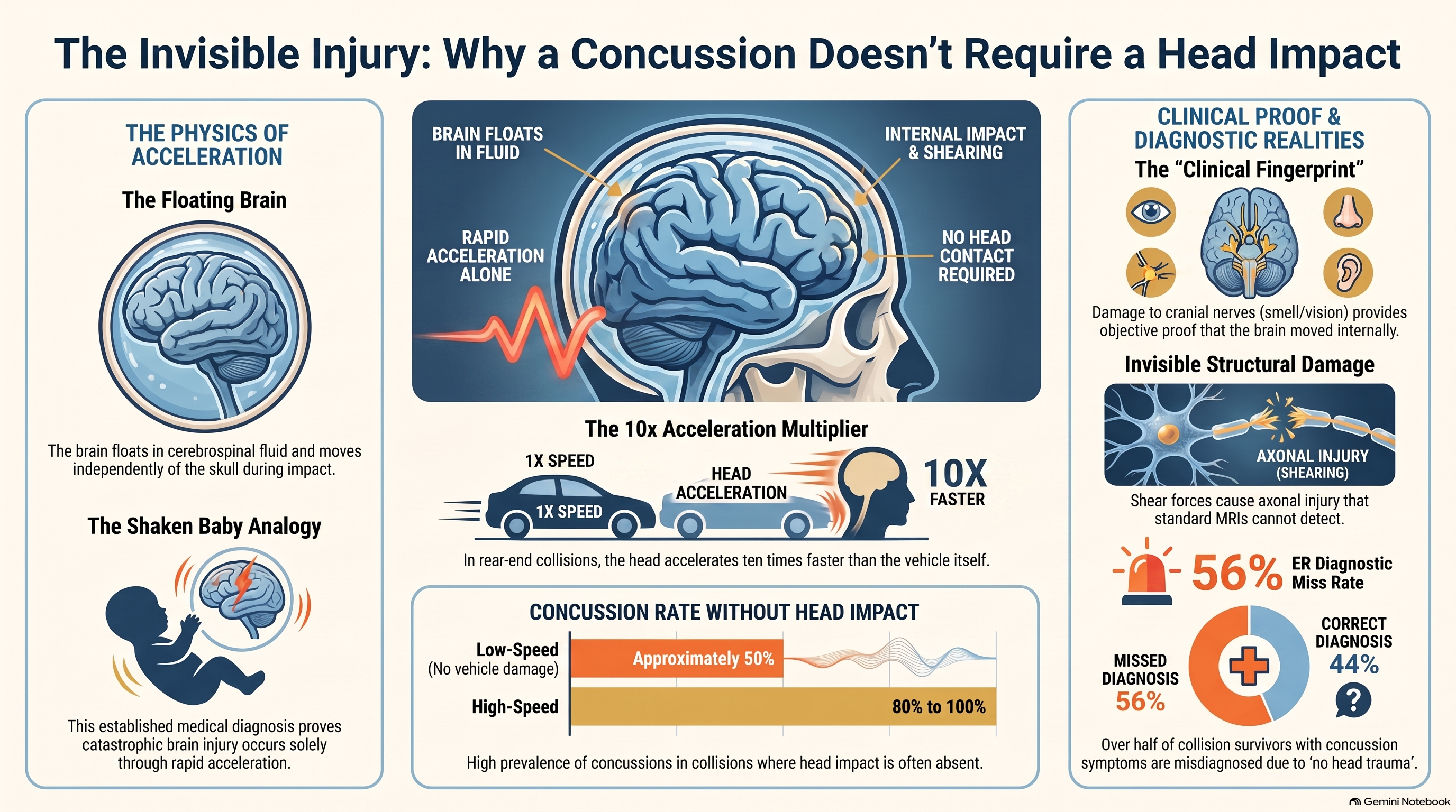
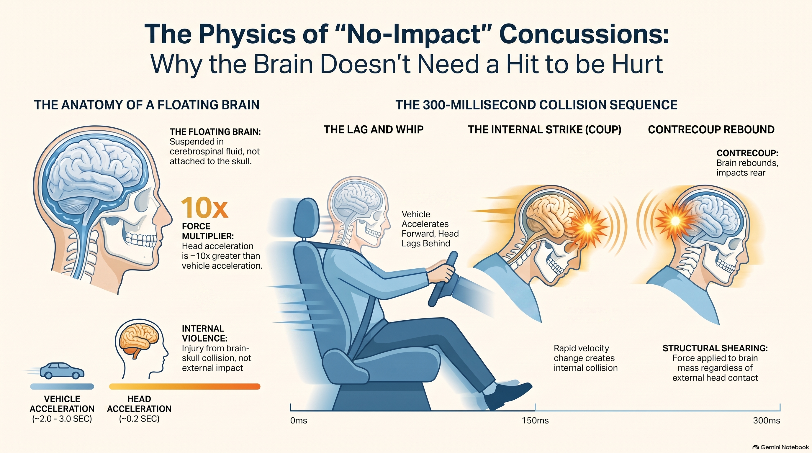
To understand why head impact is irrelevant to concussion, you need to understand one thing: the brain is not attached to the skull. It floats inside the cranium in cerebrospinal fluid, suspended and mobile. During normal movement, that floating is protective — the fluid cushions the brain against minor accelerations. During rapid acceleration-deceleration, that same mobility becomes the mechanism of injury.
When a vehicle is struck from behind, the following sequence unfolds in approximately 300 milliseconds. The vehicle accelerates forward. The seat and torso follow. The head, connected to the body by the neck’s musculature and soft tissue, lags behind — then accelerates forward in a whipping motion. The skull decelerates as the neck reaches its end range. But the brain, floating inside the skull in cerebrospinal fluid, continues moving. It impacts the interior of the skull on the side of initial motion — the coup injury — then rebounds and impacts the opposite side, the contrecoup injury.
Nowhere in this sequence is external head contact required. The injury is internal to the system: skull and brain moving at different velocities, with the brain as the delayed, mobile component. The violence happens between the brain and its own housing, not between the head and any external surface.
The physics are elementary: Force = Mass × Acceleration. The brain has mass. The collision produces acceleration. Force is applied to the brain regardless of whether anything touches the outside of the head.
There is a specific data point from the biomechanical literature that makes this concrete: in a rear-end motor vehicle collision, the head and neck experience acceleration approximately ten times greater than the vehicle itself. The vehicle may decelerate from 30 miles per hour to zero over two or three seconds. The head and neck complete their acceleration in approximately 0.2 seconds — a tenth of the time. This differential is what translates a moderate vehicle impact into a severe acceleration event for the structures inside the skull.
Shaken Baby Syndrome: The Proof That Was Already There
The scientific community has had definitive proof that brain injury occurs without head impact for decades. The mechanism is called shaken baby syndrome, and it is mechanistically identical to what happens in a motor vehicle collision.
Shaken baby syndrome produces catastrophic brain injury — subdural hemorrhage, diffuse axonal injury, retinal hemorrhage — through rapid repetitive acceleration-deceleration of the infant’s head. No impact. No contact with any external surface. Nothing touches the baby’s head from outside. The injury is produced entirely by the inertial forces created by the shaking motion, which cause the brain to move violently within the skull.
The neurological, pathological, and biomechanical research on shaken baby syndrome has been built over fifty years and includes some of the most rigorous work in pediatric neurology. Its central finding — that acceleration alone, without impact, is sufficient to produce severe traumatic brain injury — is not contested in the scientific literature. It is accepted so completely that it forms the evidentiary basis for criminal prosecution.
If acceleration alone, without any head contact, can produce catastrophic brain injury in an infant, then the question becomes: why would we assume adults require head contact for any brain injury to occur? The physics of brain motion within the skull are the same regardless of patient age. The mechanism of injury is identical. Shaken baby syndrome is not a special exception to the rules of how brains are injured — it is a demonstration of the rule.
The correct analogy for a rear-end collision patient who says “I didn’t hit my head” is not “then you probably don’t have a concussion.” The correct analogy is: the mechanism that injured your neck is the same mechanism that moved your brain inside your skull. If the force was sufficient to damage cervical ligaments — and the presence of neck pain, restricted motion, and tenderness tells us it was — the force was sufficient to produce brain acceleration. Shaken baby syndrome proves that brain acceleration alone produces brain injury. The logical chain is complete.
A useful formulation from the clinical literature: It is not possible to introduce enough force to damage cervical spine structures without also bouncing the brain inside the cranium. If the neck is injured, the brain moved. If the brain moved, a concussion occurred. The presence of a cervical injury diagnosis is, in this sense, an indirect concussion indicator — not proof in isolation, but a biomechanical premise that demands concussion screening.
What the Prevalence Data Actually Shows
The clinical literature on concussion rates in motor vehicle collisions has produced figures that should have permanently retired the “no head impact = no concussion” assumption years ago. They haven’t, largely because the figures are not well known outside specialized circles.
Concussion occurs in approximately 50% of low-speed collisions — collisions at speeds that produce no vehicle damage, no airbag deployment, and no external indication of severity. In higher-speed collisions, concussion rates range from 80% to 100%. These are not outlier figures from one or two studies. They are the consensus range across multiple research groups, study designs, and patient populations.
Independently, approximately 50% of all concussions — across sports, falls, and assault combined — occur in motor vehicle collisions. This makes motor vehicle crashes the single largest contributor to concussion in the general population, exceeding sports injuries in absolute numbers.
Most of these collision-related concussions occur without any head contact with an external surface. Head-to-steering-wheel impacts have been dramatically reduced by airbag deployment. Head-to-headrest impacts are prevented by the headrest itself. What accounts for the majority of cases is the inertial mechanism: the brain moving within the skull because the collision produced sufficient acceleration, with no external surface involved.
“56% of emergency room patients transported after a motor vehicle collision had documentation indicating concussion-consistent symptoms without ever receiving a formal concussion diagnosis.”
Research has found that 56% of emergency room patients transported after a motor vehicle collision had documentation indicating concussion-consistent symptoms without ever receiving a formal concussion diagnosis. In many of these cases, the note recorded that the patient “denied head trauma” — and the provider stopped there. The DSM-5 and CDC diagnostic criteria for concussion require exactly two things: a trauma history and at least one positive finding from a specific symptom cluster. Neither criterion mentions head impact.
Diffuse Axonal Injury: When the Damage Doesn’t Show on Standard Imaging
The neurological mechanism most relevant to understanding non-impact concussion is diffuse axonal injury — a pattern of brain injury caused by shear forces applied to axons throughout the white matter during rapid acceleration-deceleration.
White matter and gray matter have different densities and different mechanical properties. During rapid acceleration, these two tissues move at different velocities — like a two-layer system where the layers slide against each other. The shearing forces generated at the interface between gray and white matter stretch, twist, and in severe cases sever the axonal connections that run through white matter tracts, disrupting the brain’s internal communication network.
Diffuse axonal injury was historically understood as a finding in severe traumatic brain injury — the diffuse axonal disruption seen in patients with prolonged unconsciousness and poor outcomes. More recent research has substantially revised this understanding. Diffusion tensor imaging (DTI), a specialized MRI technique that maps white matter tract integrity, has made mild diffuse axonal injury visible in a population previously considered to have no structural injury: patients with mild traumatic brain injury who never lost consciousness and who present with entirely normal standard MRI findings.
Studies using DTI in mild TBI populations — including motor vehicle collision patients — consistently find reduced fractional anisotropy in white matter tracts compared to matched controls. These findings are present in the absence of any abnormality on standard MRI. They are present in patients who scored normal on initial neurological examination. And they are present in patients who never reported head contact with any surface.
The clinical implication is significant. A patient who presents after a motor vehicle collision with cognitive complaints, sleep disruption, headache, or sensitivity to light or sound, and who has a normal standard MRI, is not necessarily neurologically intact. They may have white matter changes that standard MRI cannot detect and that DTI would reveal. The absence of standard MRI findings is not evidence of neurological integrity — it is evidence of the limitations of standard MRI.
This matters because diffuse axonal injury explains the cognitive symptoms that collision patients report and that are frequently dismissed when imaging is negative. The processing speed slowing, the word-finding difficulty, the working memory deficits, the failures of sustained attention: these are the functional correlates of axonal disruption in the white matter tracts that connect and coordinate cortical processing. They are not psychological. They are not exaggeration or poor coping. They are the expected clinical presentation of a brain whose internal communication network has been mechanically disrupted by acceleration forces that never required anything to touch the outside of the head.
Research now shows that diffuse axonal injury can occur in fully awake patients who never lose consciousness and who report nothing unusual about their cognitive state at the time of the collision. The older assumption — that diffuse axonal injury is reserved for patients with extended loss of consciousness — has been replaced by a more refined understanding: the severity of DAI exists on a continuum, and its milder forms occur at the acceleration levels produced by motor vehicle collisions in patients who walked out of the emergency department without a brain injury diagnosis.
Rotational Acceleration: Why Seatbelts Don’t Eliminate the Risk
There is a specific biomechanical factor that amplifies the neurological damage produced by motor vehicle collisions significantly above what linear acceleration alone would generate: rotation.
When a seatbelted occupant is involved in a rear-end collision, the seatbelt restrains one shoulder more effectively than the other, producing a differential restraint that causes the torso to rotate as it decelerates. The head and neck, extending above the point of restraint, undergo both linear and rotational acceleration as a result. In virtually every seatbelted collision, rotation is biomechanically guaranteed.
Rotational acceleration is uniquely damaging to brain tissue. The brain is roughly spherical, and rotation produces shear forces distributed throughout the volume of the brain rather than concentrated at a single coup-contrecoup impact site. Rotational forces apply maximum shear stress precisely at the boundary between gray and white matter — the tissue interface where diffuse axonal injury originates. The brainstem, which undergoes torsional stress during head rotation, is particularly vulnerable to the stretching and twisting of axonal projections.
Research using physical models and cadaveric specimens has consistently found that rotational acceleration produces greater neurological damage per unit of force than linear acceleration alone. A 2011 study comparing injury patterns in different collision types found that rotational head acceleration was the single strongest predictor of brain injury severity — more predictive than peak linear acceleration or change in velocity.
The cranial nerves, which exit the brain through foramina in the base of the skull, are further stressed by rotational forces. During rotational head acceleration, these nerve filaments are twisted as the brain rotates within the skull base, adding a torsional component to the stretching injury they sustain from linear motion.
The practical implication is that a patient who was properly seatbelted at the time of collision was not protected from the neurological effects of the impact. The seatbelt protected them from striking the windshield or being ejected — consequences that would have been far more severe. It did not prevent their brain from undergoing rotational acceleration. Proper safety restraint and neurological injury are not mutually exclusive. They frequently coexist.
Cranial Nerve Findings: The Clinical Fingerprint of Brain Movement
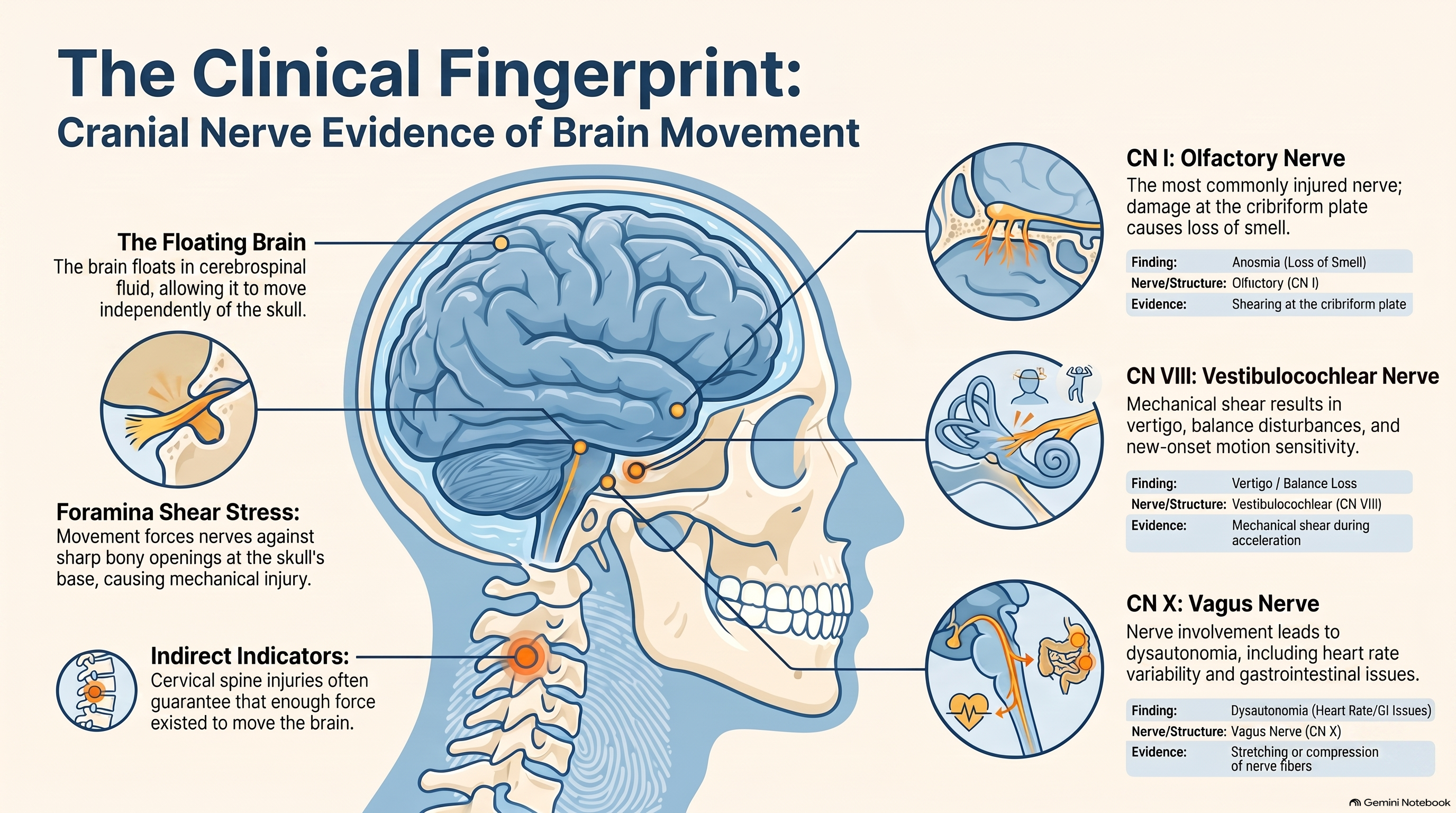
One of the most practically useful insights from the neuroscience of non-impact concussion is this: any cranial nerve finding in a post-collision patient is direct clinical evidence that the brain moved during the collision — evidence that is objective, examination-based, and not dependent on any assertion about what the patient did or did not strike their head on.
The cranial nerves exit the brain and enter the peripheral nervous system through foramina — openings — in the base of the skull. When the brain moves within the skull during acceleration, these nerves are stretched, compressed, or sheared at the point where they pass through those bony openings. The injury leaves a clinical fingerprint: dysfunction of the specific nerve that was stretched or compressed.
The most commonly injured cranial nerve in motor vehicle collisions is the olfactory nerve (CN I), which passes through the cribriform plate — a thin, perforated bony structure at the base of the anterior cranial fossa. The olfactory nerves are fine filaments that are easily damaged by the brain’s forward movement during the coup phase of acceleration. Anosmia or hyposmia — loss or reduction of smell — following a collision is, by itself, direct evidence of brain movement. It can be identified in a brief clinical examination and documented as an objective finding.
The optic nerve can be affected at the optic chiasm, where the two optic nerves cross at the base of the brain. Visual field defects in a patient who did not strike their head indicate that the brain moved with sufficient force to stress the optic system at this crossing point. Vestibular dysfunction related to the vestibulocochlear nerve (CN VIII) is another common post-collision finding, presenting as vertigo, balance disturbance, or new-onset motion sensitivity — all consistent with the nerve’s exposure to mechanical shear forces during brain acceleration.
The vagus nerve (CN X) deserves particular attention because of its role in autonomic regulation. Patients who develop dysautonomia following a collision — heart rate variability changes, orthostatic intolerance, altered gastrointestinal motility — may be presenting with the autonomic consequences of vagal nerve involvement, which in turn reflects brain movement during the collision. These presentations are rarely connected to their neurological origin in clinical settings where the history of no head contact has been accepted as a reason not to screen.
The pituitary gland sits suspended beneath the brain in the bony sella turcica. When the brain moves forward during a rear-end collision, the pituitary is driven against the posterior wall of the sella. This compression can produce pituitary dysfunction: dysregulated output affecting the adrenal axis, thyroid function, reproductive hormones, and growth hormone production. Patients who develop menstrual irregularities, patterns of fatigue consistent with adrenal axis dysregulation, or changes in their hormonal landscape following a collision without prior history of these problems may be presenting with objective evidence that the brain moved — evidence expressed in the endocrine system.
In each of these cases, the clinical finding is not merely a symptom to record. It is proof of mechanism — direct evidence, visible on examination or measurable by laboratory testing, that the physics completed as predicted regardless of the absence of external head contact.
What Proper Screening and Documentation Look Like
The DSM-5 definition of mild traumatic brain injury and the CDC’s concussion criteria share a common structure: a trauma history plus at least one positive finding from a defined symptom cluster — headache, dizziness, sleep disturbance, cognitive complaints, emotional changes, or sensory sensitivity. Not one of these symptoms requires head contact to be attributable to a collision.
A proper post-collision concussion screen does not begin with “Did you hit your head?” It begins with the mechanism, and it proceeds systematically through the symptom domains using validated tools such as the SCAT5, the Post-Concussion Symptom Scale, or the King-Devick test.
When a patient reports no head contact, the clinical record should not say “patient denies head trauma” and stop. It should accurately capture what the science says happened:
Documentation language: “Patient reports no direct contact between head and any external surface during the collision. Mechanism of injury involved rear-end impact with estimated velocity change of [X] mph, producing hyperextension-hyperflexion of the cervical spine. Per established biomechanics, this mechanism generates brain acceleration at approximately ten times the vehicle’s deceleration rate. Brain acceleration without head-to-surface contact is the mechanism of injury established in the shaken baby syndrome literature, and is consistent with diffuse axonal injury patterns documented in motor vehicle collision populations using diffusion tensor imaging. Examination reveals [specific findings]. Diagnosis: Concussion, biomechanically caused by acceleration-deceleration forces during motor vehicle collision.”
This documentation accomplishes three things. It establishes the mechanism with clinical and scientific precision. It preempts the defense argument that the absence of head impact means the absence of brain injury. And it creates a clinical record that accurately represents what the science says — not what the patient assumed before you educated them.
The patient education component matters for the same reasons. When a patient understands that their brain floats inside their skull, that the collision moved that brain regardless of what their head did or did not touch, and that the symptoms they are experiencing are the neurological consequences of that movement rather than anxiety or poor coping — they engage with their recovery differently. They stop minimizing symptoms that feel “psychological.” They report cranial nerve changes they had written off as stress. They show up to the follow-up appointments that are, for post-concussion management, the difference between identifying and treating a complication early versus managing a chronic sequela a year later.
Conclusion
The belief that concussion requires head impact is not supported by neuroscience, physics, or the diagnostic criteria published by the DSM-5 and the CDC. It is a cultural assumption that persists because it sounds intuitive, because it is rarely directly challenged at intake, and because patients themselves arrive having already accepted it as fact.
The science is clear. Concussion is an acceleration injury. The brain moves within the skull during rapid acceleration-deceleration. That movement — not any impact — is the mechanism. Shaken baby syndrome proved this without ambiguity decades ago. The whiplash prevalence data confirms it at rates between 50% and 100% depending on collision severity. The biomechanics of rotational acceleration explain why the seatbelted occupant is not protected from neurological injury. The cranial nerve findings provide the clinical fingerprint. Diffusion tensor imaging reveals the structural evidence that standard MRI cannot see.
Every patient who presents after a motor vehicle collision should be screened for concussion as a matter of clinical protocol — regardless of whether they report head contact with any surface. The screening takes minutes. The documentation takes a paragraph. What it captures is the full scope of what the collision did to the brain, not merely what the patient assumed it could not have done.
The patient who says “I didn’t hit my head, so I don’t think I have a concussion” is not offering a diagnosis. They are offering an assumption built on a misconception that the clinical community has a responsibility to correct. Your job is to replace the assumption with the science — to explain the floating brain, the shaken baby analogy, the physics of coup-contrecoup — and to document the science in terms that a reviewing physician, a defense attorney, and a jury can follow and cannot reasonably dismiss.
The brain was injured. The physics tells us so. The only question is whether anyone looked.
References
- Harmon KG, et al. American Medical Society for Sports Medicine Position Statement: Concussion in Sport. British Journal of Sports Medicine, 2013; 47(1):15–26.
- Guskiewicz KM, Mihalik JP. Biomechanics of Sport Concussion: Quest for the Elusive Injury Threshold. Exercise and Sport Sciences Reviews, 2011; 39(1):4–11.
- Johnson VE, Stewart W, Smith DH. Axonal pathology in traumatic brain injury. Experimental Neurology, 2013; 246:35–43.
- Cassidy JD, et al. Incidence, Risk Factors and Prevention of Mild Traumatic Brain Injury. Journal of Rehabilitation Medicine, 2004; Suppl 43:28–60.
- Sturzenegger M, et al. Presenting symptoms and signs after whiplash injury: the influence of accident mechanisms. Neurology, 1994; 44(4):688–693.
- American Psychiatric Association. Diagnostic and Statistical Manual of Mental Disorders, 5th ed. (DSM-5). Washington, DC: APA; 2013.
- Centers for Disease Control and Prevention. Traumatic Brain Injury & Concussion: Symptoms of Mild TBI and Concussion. CDC, 2023.