Not Broken, But Disconnected: How Whiplash Disrupts the Body-Brain Communication System
Within 300 milliseconds of a rear-end collision, before the driver has registered what happened, the deep cervical muscles have already contracted, stretched, and in many cases sustained micro-tears that will not appear on any X-ray. The bones may be intact. The discs may be intact. The standard imaging report will say “no acute findings.” And the patient — often sitting in front of a clinician weeks later — will be told, explicitly or implicitly, that there is nothing wrong.
There is something wrong. It just isn’t what imaging is designed to find.
In a substantial proportion of whiplash cases, the primary injury is not structural. It is informational. The tissues that govern how your body communicates its position to your brain — a system called proprioception — have been disrupted at a level that doesn’t register on standard MRI, that doesn’t produce visible swelling, and that can persist for months or years without any external marker of its severity. The muscles aren’t broken. The signal they’re sending is.
Understanding this distinction is not a semantic exercise. It changes what treatment looks like, what recovery requires, and why patients who are told they “should be better by now” often are not — and why the problem, left uncorrected, can become structural over time.
The Signal System Your Spine Depends On
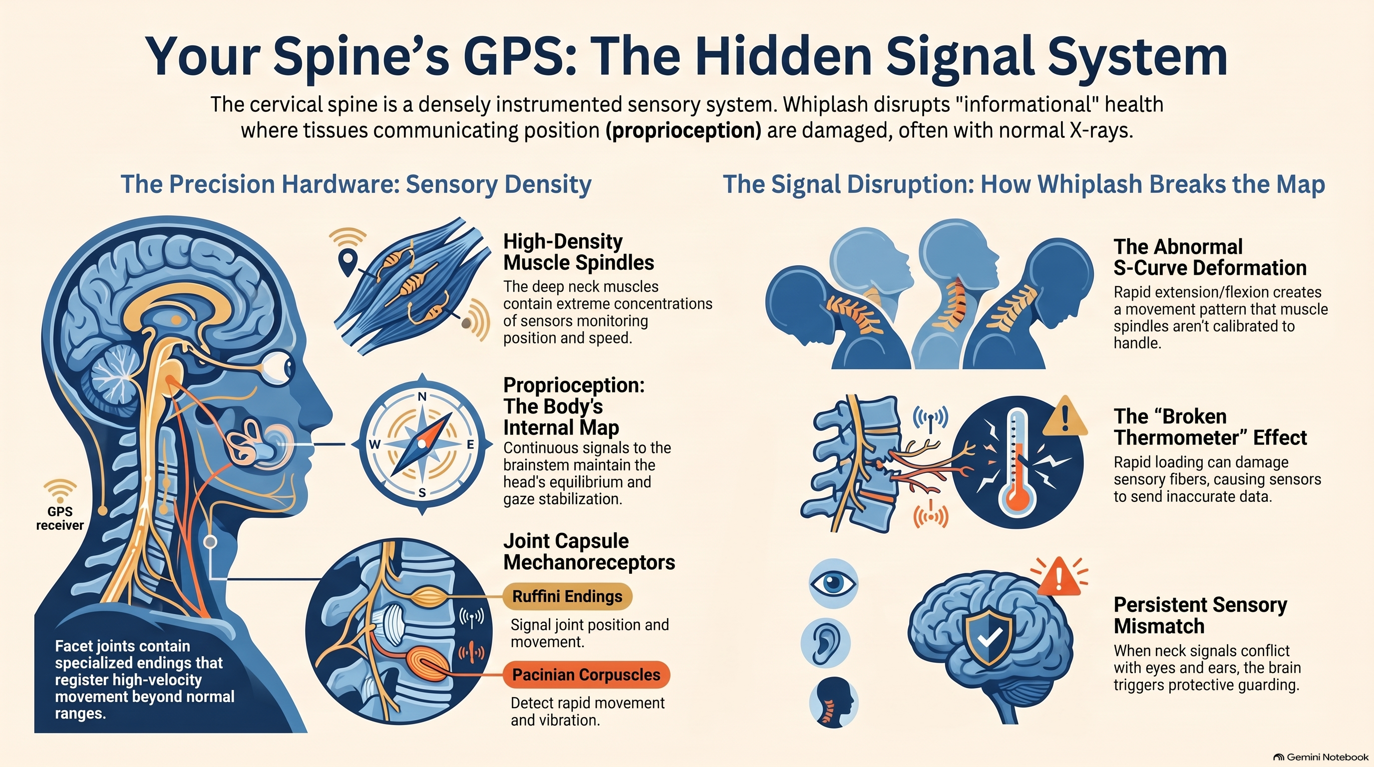
The cervical spine is not just a column of bones and discs. It is one of the most densely instrumented sensory structures in the human body.
Embedded in the deep cervical muscles — particularly the multifidus and semispinalis — are mechanoreceptors called muscle spindles. These spindles monitor muscle length and the rate at which length is changing, sending continuous signals to the brainstem and cerebellum about the position, speed, and orientation of the head and neck in space. This system is called proprioception: the body’s internal sense of where it is.
The density of muscle spindles in the cervical muscles is not an accident of anatomy. The head is approximately 10–12 pounds, sits atop a highly mobile seven-vertebra column, and must be held in dynamic equilibrium while the eyes track moving objects and the vestibular system monitors balance. The precision required for this coordination depends on continuous, accurate proprioceptive input from the cervical musculature. Disruption of that input disrupts everything downstream.
The facet joint capsules — the fibrous tissue surrounding the small joints between vertebrae — also contain a dense concentration of mechanoreceptors, including Ruffini endings, Pacinian corpuscles, and free nerve endings. These contribute additional proprioceptive data about joint position and movement. In a rear-end collision, the facet joints undergo rapid stretching beyond their normal physiological range. The mechanoreceptors within them register this as a high-velocity, high-amplitude event — and they don’t always recover their baseline function afterward.
The cervical spine’s role in postural control and gaze stabilization is coordinated through the cervicocollic reflex and the cervico-ocular reflex — two automatic systems that depend on accurate, real-time proprioceptive input from the neck. When that input is distorted, the reflexes misfire. The results show up in the clinic as gaze instability, difficulty tracking moving objects, dizziness with head movement, and postural sway — symptoms that, in the absence of vestibular pathology or intracranial injury, often have their root in the disrupted sensory output of injured cervical muscle spindles.
What Whiplash Does to the Signal
The acceleration-deceleration injury of a rear-end collision creates a specific pattern of tissue stress that targets the proprioceptive system with unusual precision.
During the collision, the cervical spine undergoes a characteristic S-curve deformation: the lower cervical segments rapidly extend while the upper segments flex, creating a temporary shape that does not occur in normal voluntary movement. This motion pattern is beyond the range that the cervical muscles normally encounter during daily activity, which means it occurs in a zone where muscle spindle calibration is not routinely maintained. The spindles are stretched in a pattern they were not designed to handle.
Research by Sterling and colleagues has demonstrated that whiplash injury produces measurable changes in cervical proprioception that persist well beyond the acute phase of recovery. Using validated clinical tools — the joint position error test, cervical range of motion measurement, and proprioceptive matching tasks — studies have consistently found that patients with whiplash-associated disorders show significantly higher joint position error than matched controls. This means that when they close their eyes, move their head away from neutral, and attempt to return to the starting position, they miss — by a margin that reflects the degree of sensory disruption.
The implications of this finding extend beyond a clinical curiosity. Joint position error is a measurable proxy for the accuracy of the proprioceptive signal. When that signal is inaccurate, the brain’s internal model of where the head and neck are in space — its map of the body — becomes incorrect. The brain receives data that doesn’t match reality. It compensates. Sometimes it compensates visibly, through altered posture and movement patterns. Sometimes it compensates internally, through increased muscle guarding, heightened neural sensitivity at the spinal cord level, and an upregulated threat response.
A 2006 study by Treleaven and colleagues found that dizziness — one of the most commonly reported and frequently dismissed symptoms following whiplash — was significantly associated with impaired cervical proprioception. The dizziness was not vestibular in origin. It originated in the disrupted sensory output of the cervical musculature, which failed to provide the accurate positional data the brainstem required for balance coordination. Treating the vestibular system in these patients produced limited results. Addressing the proprioceptive deficit produced meaningful improvement.
The muscle spindles themselves can be mechanically altered by the injury. Rapid eccentric loading — the type of force applied to the cervical extensors as the head whips forward — has been shown to produce intrafusal muscle fiber damage, meaning the spindle structure itself is injured. This is not soft tissue bruising. It is damage to the sensory apparatus that monitors muscle length. A thermometer with a broken sensor doesn’t give accurate temperature readings. A muscle spindle with damaged intrafusal fibers doesn’t give accurate position data.
“The muscles aren’t broken. The signal they’re sending is.”
When the Map No Longer Matches the Territory
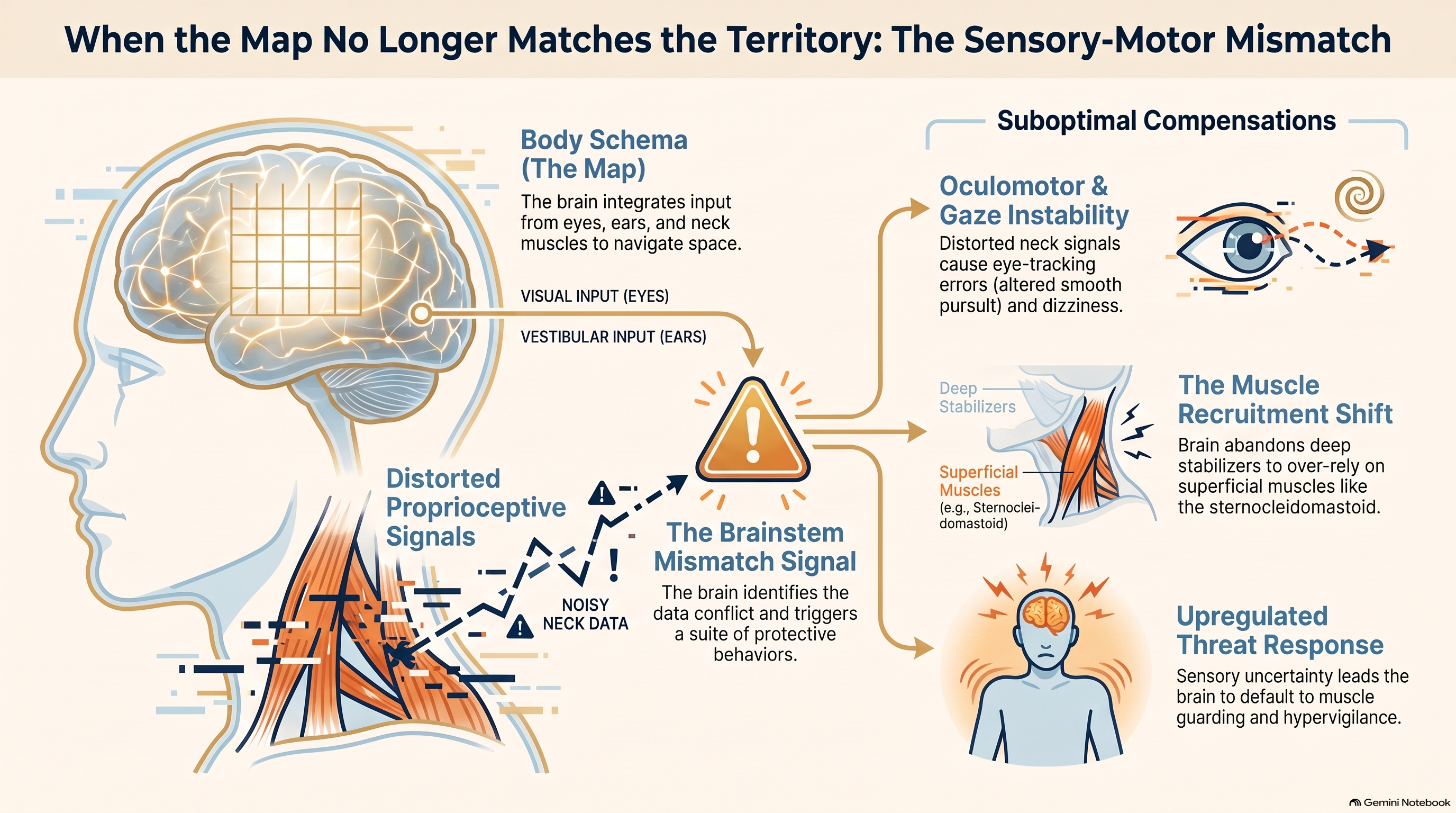
The brain constructs a real-time model of the body’s position in space — sometimes called a body schema — by integrating proprioceptive input from muscles and joints, vestibular input from the inner ear, and visual input from the eyes. Under normal conditions, these three streams of information are consistent with each other. When they conflict, the brain generates a mismatch signal — and in the presence of injury, that mismatch can become chronic.
After whiplash, the proprioceptive signal from the cervical musculature is degraded. The vestibular and visual signals remain intact. This creates a persistent sensory conflict: the body’s joint position sense says one thing; the eyes and inner ear say another. The brainstem, which must reconcile these inputs in real time, responds to the conflict with a suite of protective behaviors — increased muscle tone to stabilize the uncertain region, heightened sensitivity to movement in the neck, and in some patients, a pattern of hypervigilance toward head and neck position that becomes its own generator of symptoms.
This mechanism is not hypothetical. Treleaven’s research group has documented that patients with chronic whiplash-associated disorders show altered smooth pursuit — the ability to track a slowly moving visual target with the eyes — at a rate significantly higher than controls or patients with acute whiplash who recovered normally. Smooth pursuit eye movement is controlled by a system that integrates vestibular, visual, and proprioceptive input. When the proprioceptive input is chronically distorted, the smooth pursuit system loses its calibration. The result is detectable on examination and measurable on specialized eye-tracking equipment.
The postural consequences are equally concrete. Research by Falla and colleagues has demonstrated that whiplash injury is associated with a shift in cervical muscle recruitment: the deep stabilizing muscles — the longus colli and longus capitis — reduce their activity, while the superficial muscles — the sternocleidomastoid and scalenes — increase theirs. This shift represents an adaptive response to the perceived instability signaled by degraded proprioceptive input. The brain, unsure of the neck’s position from moment to moment, defaults to recruiting the larger, more powerful surface muscles to maintain a gross sense of control. The result is a muscle use pattern that is less efficient, more fatiguing, and ultimately less stable than the pattern it replaced.
This recruitment shift is not simply a consequence of pain. Falla’s group showed that it persists even during tasks that do not provoke pain in patients with chronic neck pain — suggesting that it is driven by the underlying sensory disruption, not by pain avoidance alone. The muscle pattern has become reorganized at a central level. The brain has relearned how to hold up the head, and what it has learned is suboptimal.
Kinesiophobia: How Disrupted Signals Create Fear of Movement
The relationship between sensory disruption and movement fear in whiplash is not straightforward, but it is well-documented.
When the brain receives inaccurate or conflicting proprioceptive information, it cannot confidently predict the outcome of movement. Unpredictable movement outcomes trigger threat responses. The body’s threat appraisal system — centered in the amygdala and integrated through descending inhibitory and facilitatory pathways from the brainstem — interprets the sensory uncertainty as potential danger. The result, in some patients, is a progressive avoidance of movement: kinesiophobia.
Kinesiophobia — literally, fear of movement — is not a psychological fragility. It is a rational response to a proprioceptive system that is sending unreliable data. When the brain cannot accurately predict the consequences of turning the head, the safest strategy is not to turn the head. This strategy reduces the immediate threat signal. It also eliminates the proprioceptive input that would, over time, recalibrate the sensory system and reduce the mismatch. Avoidance of movement is self-perpetuating.
Key research finding: A 2008 systematic review by Söderlund and Lindberg found that kinesiophobia, assessed early in the recovery process, predicted long-term disability with greater accuracy than the severity of the initial injury, the patient’s pain intensity, or the degree of structural damage identified on imaging.
Research using the Tampa Scale of Kinesiophobia has consistently found elevated kinesiophobia scores in patients with chronic whiplash-associated disorders, and multiple studies have identified kinesiophobia as one of the strongest predictors of chronic pain and disability following whiplash injury. A 2008 systematic review by Söderlund and Lindberg found that kinesiophobia, assessed early in the recovery process, predicted long-term disability with greater accuracy than the severity of the initial injury, the patient’s pain intensity, or the degree of structural damage identified on imaging.
This finding has profound clinical implications. It means that the patient’s relationship with movement — their sense of safety or danger in activating the injured region — is more predictive of outcome than the injury itself. And it means that any treatment approach that reinforces the idea that the neck is fragile, that movement is dangerous, or that rest is protective is, in a very real sense, making the prognosis worse. The threat response requires a corrective experience, not a confirmation of threat.
The pain neuroscience education literature is relevant here. Research by Louw, Nijs, and colleagues has demonstrated that patients who receive education about the mechanisms of pain — specifically, about the role of central sensitization, threat appraisal, and the nervous system’s role in amplifying or dampening pain signals — report reduced pain intensity, increased movement tolerance, and improved function following whiplash and other musculoskeletal injuries. The education does not cure the proprioceptive deficit. But it removes the additional layer of threat that fear of movement superimposes on the underlying sensory disruption, creating space for the movement-based interventions that can begin to recalibrate the system.
Why “Rest and Wait” Is the Wrong Prescription
For decades, the standard clinical advice following whiplash was some version of rest, a soft collar, and watchful waiting. The evidence has now unequivocally reversed this guidance — and understanding why requires understanding what rest does to the proprioceptive system.
Proprioception is not a fixed capacity. It is a trained capacity. The muscle spindles that monitor cervical position are maintained and calibrated by movement — specifically, by the varied, multi-directional, unpredictable movement that characterizes daily life. When movement is restricted — by a soft collar, by protective guarding, or by pain-driven avoidance — the spindles lose the routine calibration inputs that keep them accurate. A proprioceptive system deprived of movement inputs does not recover. It drifts further from accuracy.
A landmark study compared outcomes in whiplash patients given active exercise programs versus those given rest and collar immobilization. At six months and twelve months follow-up, patients in the active exercise group had significantly better cervical range of motion, lower pain scores, and faster return to work than those in the rest group. The structural injury was not different between the groups. The treatment approach was.
The Québec Task Force on Whiplash-Associated Disorders — whose 1995 report became one of the most influential documents in whiplash management — found that early mobilization and active exercise were strongly associated with better outcomes, while passive treatments and immobilization were associated with worse ones. Subsequent research has refined and reinforced this finding across multiple healthcare systems, injury severity levels, and patient populations.
The reason immobilization produces worse outcomes is not simply that movement is “good for you.” It is that the proprioceptive system requires movement input to recalibrate the sensory-motor mismatch that the injury created. Rest preserves the mismatch. Movement, introduced gradually and purposefully, gives the brainstem and cerebellum the corrective sensory data they need to update the body schema. This is why active exercise is not just a component of whiplash rehabilitation — it is the mechanism through which rehabilitation works.
Soft cervical collars, still commonly prescribed in emergency departments, compound this problem. By restricting neck movement, they not only prevent proprioceptive recalibration but actively reinforce the message that the neck is injured and vulnerable — a message the brain’s threat appraisal system receives and amplifies. Patients who wear soft collars consistently show worse outcomes on measures of pain, disability, and return to function than those who do not. The collar is not protecting the neck. It is protecting the injury.
Restoring the Signal: What Evidence-Based Recovery Actually Looks Like
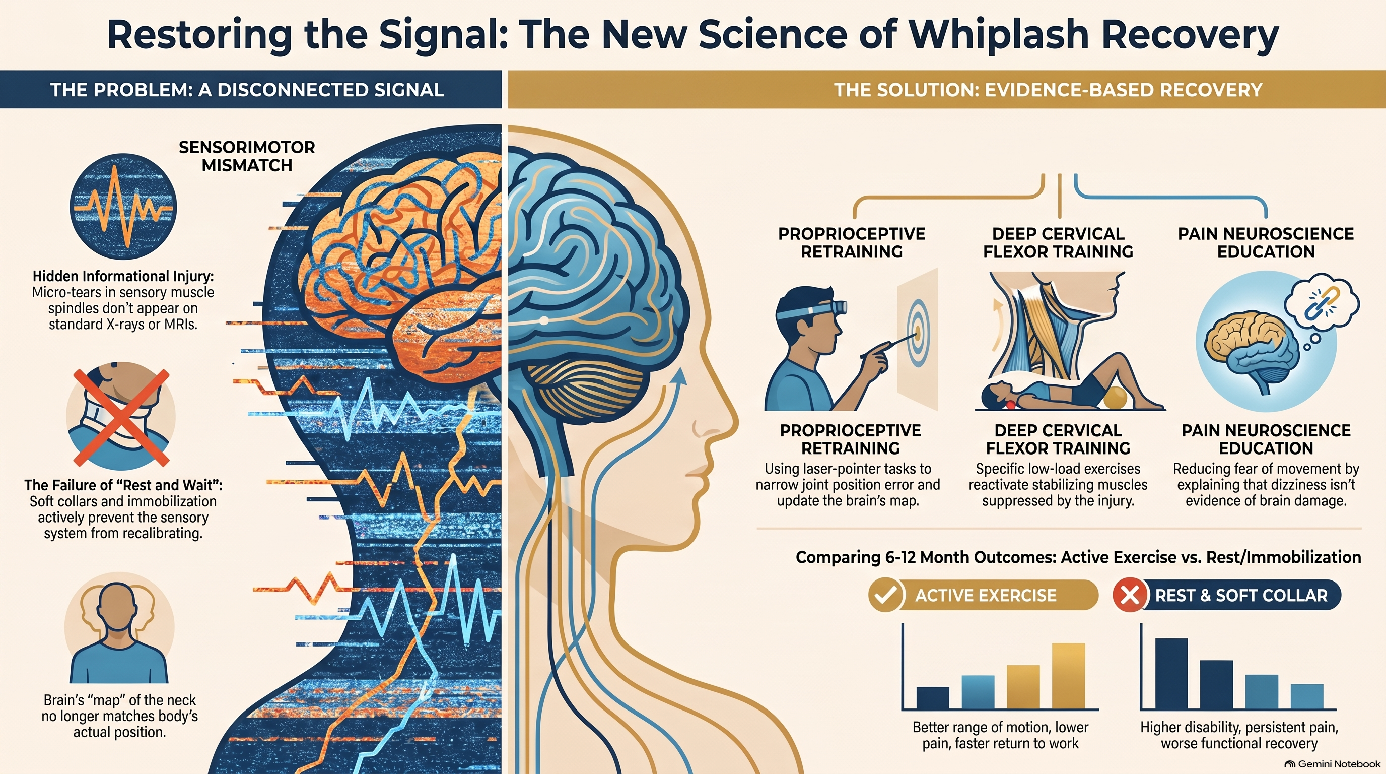
Restoring proprioceptive function after whiplash is a process of graduated sensory challenge, delivered within a framework of education and reassurance. The science is specific about what works — and it is different from what most patients expect.
Cervical proprioceptive retraining is the most directly targeted intervention. These exercises use laser pointer head repositioning tasks, head movement accuracy challenges, and oculomotor training to systematically challenge and correct joint position error. A patient places a small laser pointer on their head, moves away from a target on the wall, and returns to it with eyes closed — then measures the error. Repeated over sessions, with feedback, the brain recalibrates the relationship between motor command and sensory outcome. The mismatch narrows.
Research by Jull and colleagues — who have conducted some of the most rigorous work on whiplash rehabilitation — has demonstrated that cervical proprioceptive retraining combined with therapeutic exercise produces superior outcomes to either intervention alone, and both outperform general advice and passive treatment. The proprioceptive component is not an add-on. It targets the underlying mechanism.
Deep cervical flexor training addresses the muscle recruitment reorganization that follows whiplash injury. The Cranio-Cervical Flexion Test (CCFT) — a low-load isometric exercise performed in supine — reactivates the longus colli and longus capitis while minimizing superficial muscle dominance. Its value lies in specificity: it recruits the muscles whose recruitment has been suppressed by the injury-driven reorganization of the motor system. Multiple randomized controlled trials have demonstrated its efficacy in reducing pain and improving function in patients with neck pain, including post-whiplash presentations.
Pain neuroscience education (PNE) prepares patients to engage with these exercises by changing their understanding of what their symptoms mean. When a patient understands that their dizziness is not evidence of brain injury, that their neck tightness is not evidence of structural instability, and that their pain response is amplified by a threat-detection system that has been miscalibrated by sensory disruption — rather than by ongoing tissue damage — their kinesiophobia decreases and their willingness to engage with movement-based treatment increases.
Graded exposure to movement is the behavioral component that translates neuroscience education into functional change. Working from the patient’s specific movement fears and avoidances, the clinician constructs a hierarchy of activities from least to most threatening and guides the patient through them in order. Each successful experience contributes to the downregulation of the threat response that kinesiophobia has built.
Manual therapy has a supporting role in whiplash rehabilitation — specifically in reducing pain in the acute and subacute phases to a level that allows the proprioceptive and exercise-based work to proceed. The evidence does not support passive manual therapy as a primary or standalone treatment. But it does support its use as a facilitator of active rehabilitation — reducing the protective guarding that limits participation in the exercises that drive recovery.
Recovery timeline: Studies tracking joint position error in recovering whiplash patients have found that improvement occurs over weeks to months, with the steepest gains in patients who engaged consistently with cervical repositioning exercises and showed the lowest levels of kinesiophobia at baseline. Early identification and early movement are the two variables that most reliably alter the trajectory.
Conclusion
The standard framing of whiplash injury — as a problem of damaged soft tissue requiring rest, pain management, and time — fails to account for the most important mechanism in the majority of cases. The tissues may be injured. But the more consequential disruption is in the information system that those tissues support: the proprioceptive network that tells the brain where the neck is, how it is moving, and what the body should do next.
When that network is disrupted, the consequences cascade outward — through postural reorganization, gaze instability, dizziness, muscle guarding, and the progressive development of kinesiophobia that transforms an acute injury into a chronic disability. These consequences are measurable, addressable, and reversible — but only if the treating clinician recognizes them as the primary problem and selects interventions that target the sensory-motor mismatch, not the tissue alone.
The tissues in most whiplash cases are not beyond repair. The signal is distorted. Restoring it — through proprioceptive retraining, deep cervical muscle activation, pain neuroscience education, and graded movement exposure — is the clinical work that determines whether a patient recovers in weeks or suffers for years. The question is not whether the neck is broken. The question is whether anyone is listening to what it’s trying to say.
References
- Sterling M, Jull G, Vicenzino B, Kenardy J, Darnell R. Development of motor system dysfunction following whiplash injury. Pain. 2003;103(1–2):65–73.
- Treleaven J, Jull G, Sterling M. Dizziness and unsteadiness following whiplash injury: characteristic features and relationship with cervical joint position error. Journal of Rehabilitation Medicine. 2003;35(1):36–43.
- Falla D, Jull G, Hodges P. Feedforward activity of the cervical flexor muscles during voluntary arm movements is delayed in chronic neck pain. Experimental Brain Research. 2004;157(1):43–48.
- Söderlund A, Lindberg P. Whiplash-associated disorders — predicting disability from pain, cognition, neurology, and locomotor function. Disability and Rehabilitation. 2003;25(20):1125–1132.
- Louw A, Diener I, Butler DS, Puentedura EJ. The effect of neuroscience education on pain, disability, anxiety, and stress in chronic musculoskeletal pain. Archives of Physical Medicine and Rehabilitation. 2011;92(12):2041–2056.
- Spitzer WO, Skovron ML, Salmi LR, et al. Scientific monograph of the Quebec Task Force on Whiplash-Associated Disorders. Spine. 1995;20(8 Suppl):1S–73S.
- Jull G, Treleaven J, Versace G. Manual therapy to ease cervical joint stiffness and pain sensitization. Journal of Manipulative and Physiological Therapeutics. 2007;30(6):442–449.