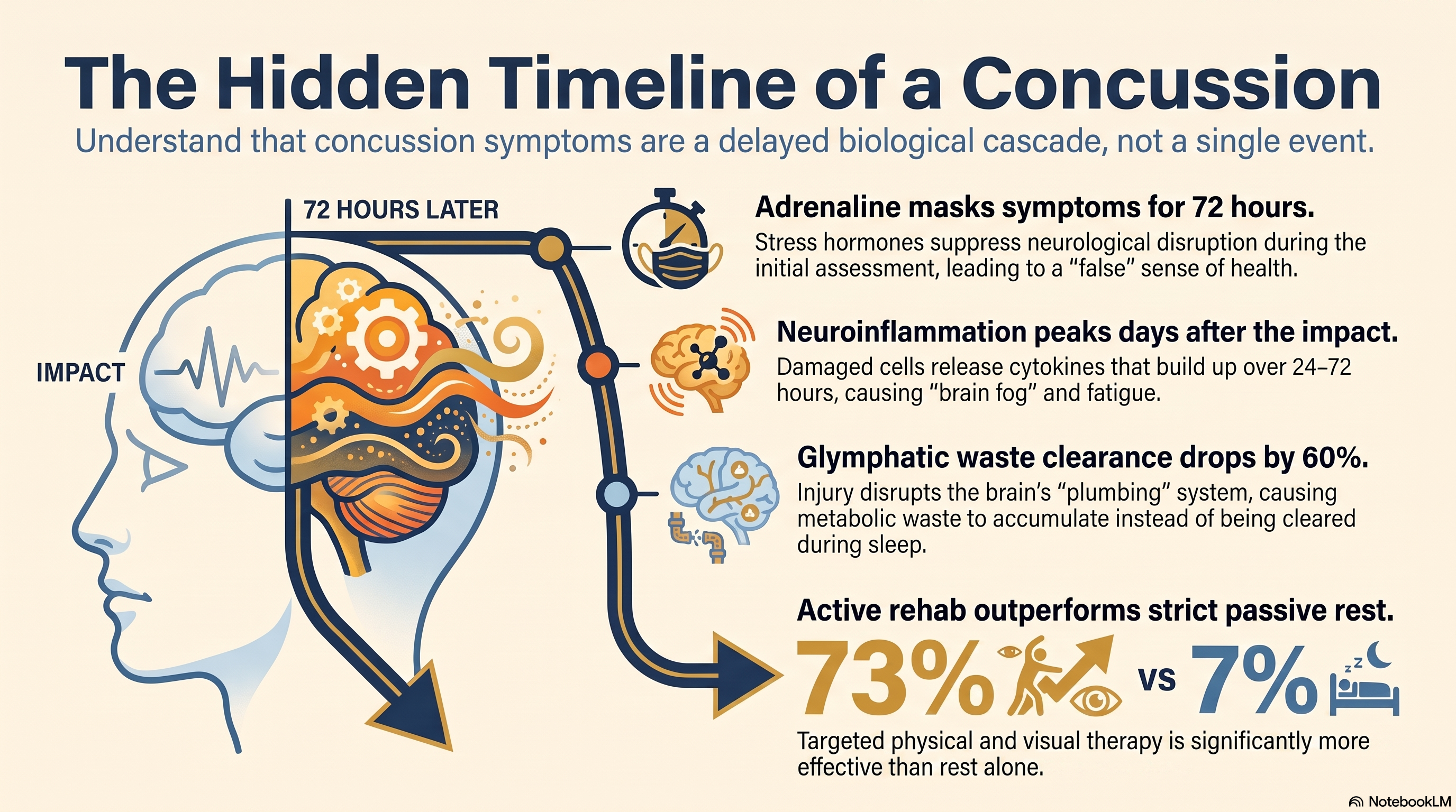
Approximately 30% of people diagnosed with concussion will still have symptoms at one month. Half of those will still have them at three months. Most of them were told the same thing in the emergency room: rest, avoid screens, return if symptoms worsen. None of them were told the symptoms hadn't started yet.
This is not a failure of communication. It is a failure of the dominant model — the idea that a concussion is an event that happens and then resolves, that the biology peaks at impact and retreats from there. The science does not support that model. What the science shows is a cascade: a neurobiological sequence that can unfold over days to weeks, shaped by adrenaline, neuroinflammation, metabolic crisis, and glymphatic dysfunction — processes that are invisible on CT scans and easy to dismiss on discharge paperwork.
Understanding why concussion symptoms appear late is not an academic exercise. It is the clinical and legal foundation for why patients deserve a different standard of care — one that anticipates delayed presentation, monitors it systematically, and treats it with an integrated rehabilitation approach rather than a prescription for passive rest.
Why the ER Misses What’s Coming
The emergency department assessment for concussion is designed to rule out what is immediately dangerous: intracranial hemorrhage, skull fracture, herniation. CT imaging is excellent at identifying those catastrophic findings. It is nearly useless for identifying the functional injury that defines most concussions.
The brain does not bleed after most concussions. It shears. The forces transmitted through the skull during a motor vehicle collision, a fall, or a blow to the head stretch and distort axons — the long fiber tracts that connect neurons and carry signals across the brain. This stretch injury, called diffuse axonal injury (DAI) at its most severe, occurs at the microscopic level. Standard CT and even most MRI protocols cannot resolve it. A normal scan is not a clean bill of neurological health. It is a statement about the resolution limits of the instrument.
“A normal CT scan is not evidence that the injury was minor. It is evidence that the assessment occurred during the window in which the injury was most likely to be underestimated.”
So the patient is discharged. The scan is normal. The headache is mild — sometimes absent entirely — and the adrenaline of the accident is still circulating. They feel, if not fine, at least functional. They go home. Three days later, they cannot get out of bed.
Adrenaline and the Masked Presentation
The role of catecholamines — adrenaline (epinephrine) and noradrenaline (norepinephrine) — in masking acute concussion symptoms is one of the most under-discussed mechanisms in post-injury education.
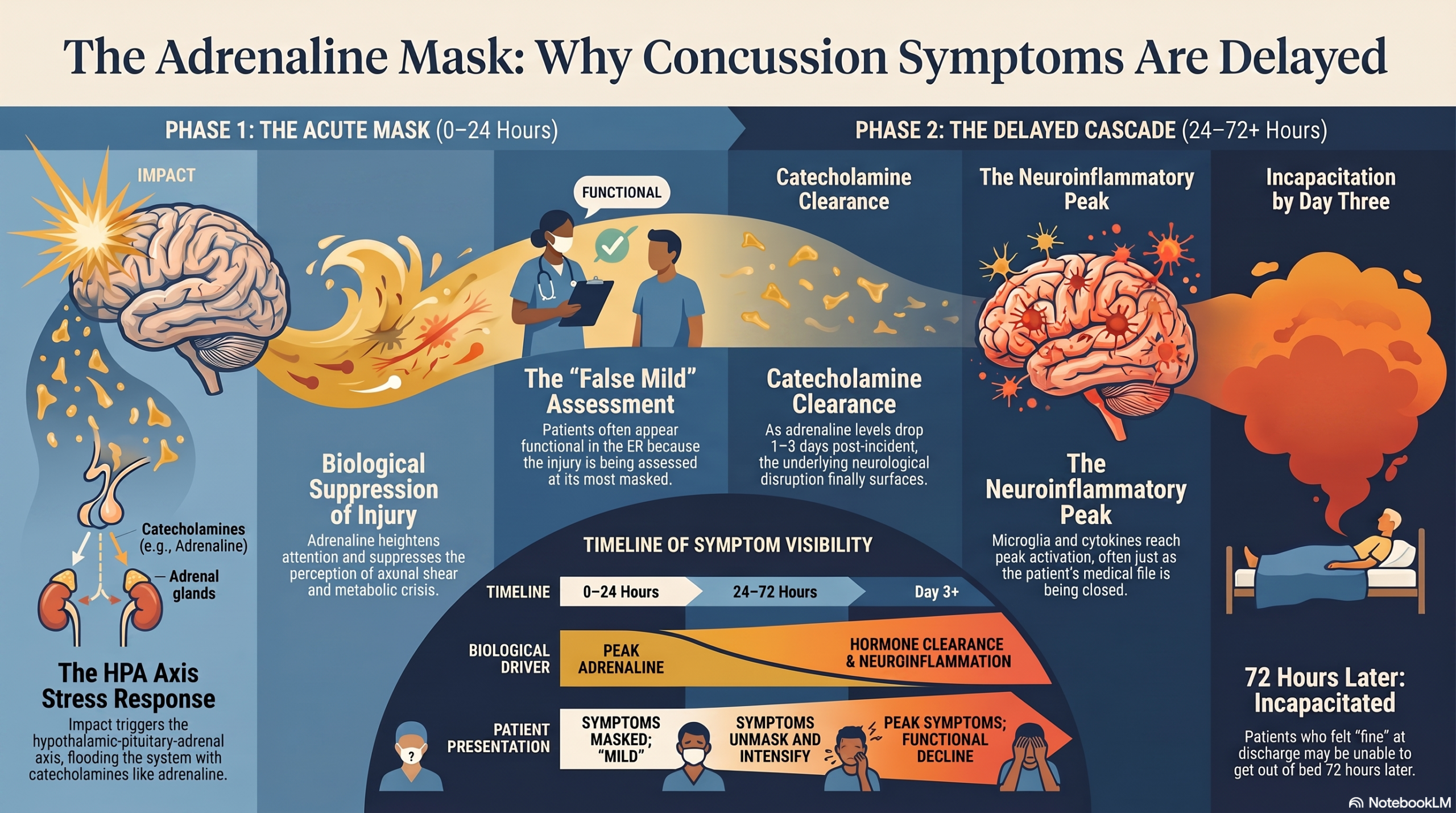
At the moment of trauma, the hypothalamic-pituitary-adrenal axis triggers an immediate stress response. Cortisol and catecholamines flood the system. Heart rate rises. Blood pressure increases. Attention narrows. The neurological disruption that has just occurred is effectively suppressed — not healed, but suppressed — by the same biological machinery that evolved to keep an injured organism functional long enough to survive.
This is protective in the short term. It becomes diagnostically treacherous in the emergency room, where the assessment is occurring during the acute sympathetic surge. Patients report fewer symptoms than they will report 48 to 72 hours later, not because they are exaggerating subsequently, but because the neurological disruption was masked at the time of assessment.
Research by Pertab et al. (2009) documented this phenomenon, noting that the acute neuropsychological presentation often underestimates the severity of injury in the first hours post-impact. The same principle applies to any high-stress trauma context — including motor vehicle collisions, where the shock of the event itself generates a prolonged sympathetic state.
By the time the adrenaline clears — typically 24 to 72 hours post-incident — the underlying neurobiological injury is no longer masked. It is also no longer being evaluated. The patient is home. The file is closed.
The Neuroinflammatory Cascade
Concussion does not injure the brain in the way a broken bone injures a limb. It triggers a cascade. And like all biological cascades, the downstream effects can be more disabling than the initial event.
Within hours of the traumatic force, damaged neurons and supporting glial cells release inflammatory cytokines — signaling molecules that recruit immune cells to the injury site. In a contained peripheral injury, this response is efficient and self-limiting. In the brain, it is structurally complicated by the blood-brain barrier and by the unique immunological environment of the central nervous system.
Microglia — the brain’s resident immune cells — become activated. In moderate quantities and for limited durations, this activation is appropriate: it clears cellular debris and initiates repair. But research published in the Journal of Neuroinflammation has documented that microglial activation after concussive injury can persist for months to years, well beyond the period of acute injury, and that this chronic low-grade neuroinflammation contributes to the symptom profile patients experience as post-concussive syndrome (Ziebell & Morganti-Kossmann, 2010).
The neuroinflammatory state produces specific symptoms: cognitive fog (difficulty with concentration, memory encoding, and processing speed), fatigue, sleep disruption, light and noise sensitivity, and mood dysregulation. These are not psychological reactions to being injured. They are physiological outputs of an ongoing neuroinflammatory process that has a documented time course.
Importantly, that time course does not peak on day one. Cytokine production and microglial activation build over the first 24 to 72 hours and can remain elevated for weeks. The patient who felt adequately functional on the day of the ER visit may be experiencing peak neuroinflammation by the time their follow-up appointment occurs — if one was scheduled at all.
Glymphatic Dysfunction and the Sleep–Concussion Connection
One of the most important discoveries in modern neuroscience — and one of the most underutilized in clinical concussion management — is the glymphatic system.
Described in landmark research by Maiken Nedergaard’s group at the University of Rochester (Iliff et al., 2013), the glymphatic system is the brain’s waste-clearance network. It operates primarily during deep slow-wave sleep, using cerebrospinal fluid (CSF) to flush metabolic byproducts — including amyloid-beta and tau proteins — out of the brain’s interstitial space and into the venous circulation for elimination.
Traumatic brain injury, even at the mild concussive level, disrupts glymphatic function. Aquaporin-4 (AQP4) channels — the water channels on astrocytes that drive glymphatic flow — are structurally damaged by the shear forces of concussion. Research by Iliff et al. (2014) demonstrated that mice with TBI showed a 60% reduction in glymphatic transport capacity in the weeks following injury. The downstream consequence is accumulation of metabolic waste products in the brain tissue — waste that, under normal circumstances, would have been cleared during sleep.
“The injury impairs the very system responsible for clearing the biological evidence of injury. The brain’s repair process is compromised by the injury itself.”
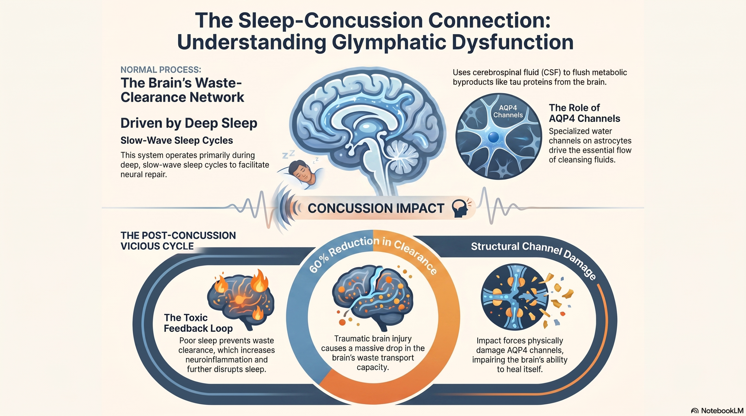
This creates a vicious cycle that explains why concussion patients so often report that their symptoms worsen over time rather than resolve. Sleep disturbance — which affects the majority of concussion patients — compounds this further. Less deep sleep means less glymphatic clearance, which means more metabolic waste accumulation, which means more neuroinflammation, which means worse sleep. The patient who “just can’t sleep right” after a concussion is experiencing a physiologically coherent failure of neural repair, not anxiety or poor sleep hygiene.
The Rest-Only Protocol and Its Evidence Problem
For decades, the standard concussion management prescription was cocoon therapy: strict physical and cognitive rest until symptom resolution. No screens, no reading, no exercise, minimal conversation. The rationale was intuitive — an injured brain needs rest to recover.
The evidence base for this approach has not held up.
A landmark randomized controlled trial by Thomas et al. (2015), published in Pediatrics, assigned concussed patients to either strict rest or usual care. The strict rest group did not recover faster. In fact, they reported higher rates of persistent symptoms at follow-up. A subsequent Cochrane review on the subject concluded that there was insufficient evidence to support extended rest and emerging evidence that it may be harmful.
Prolonged rest does not address the cervicogenic and vestibular components of post-concussive symptoms — components that, if left untreated, become self-reinforcing sources of symptom perpetuation entirely independent of the original brain injury.
Why would rest prolong symptoms? Several mechanisms have been proposed. Physical inactivity allows the glymphatic system — which is partly driven by the interstitial pressure dynamics of movement and cardiovascular flow — to operate below optimal capacity. Cognitive rest, extended beyond the first 24 to 48 hours, may impair the neural reconnection and neuroplastic adaptation that is necessary for functional recovery. Social isolation, often a byproduct of cocoon therapy, is itself associated with worse neurological outcomes.
More specifically, prolonged rest does not address the cervicogenic and vestibular components of post-concussive symptoms — components that, if left untreated, become self-reinforcing sources of symptom perpetuation entirely independent of the original brain injury.
Cervicovestibular and Visual Rehabilitation: The Evidence-Based Path
The cervical spine does not survive a concussive event neutrally. The same forces that cause the brain to move inside the skull — sudden acceleration, deceleration, rotational dynamics — simultaneously transmit mechanical stress through the cervical musculature, facet joints, and proprioceptive afferents of the upper cervical spine.
Cervicogenic dizziness, cervicogenic headache, and impaired cervical proprioception are common sequelae of concussion that are frequently attributed to the brain injury itself, when in reality they have a significant musculoskeletal component that is both diagnosable and treatable. Research by Schneider et al. (2014) demonstrated that concussion patients with cervicogenic and vestibular dysfunction recovered significantly faster when treated with targeted cervical and vestibular physiotherapy compared to rest alone — with recovery rates of 73% versus 7% achieving full clearance at eight weeks.
That is not a marginal difference. It is a categorical one.
The vestibular system — comprising the inner ear apparatus, its central processing pathways in the brainstem and cerebellum, and its projections to the visual and proprioceptive systems — is exquisitely vulnerable to concussive injury. Benign paroxysmal positional vertigo (BPPV), which involves dislodged otoconia in the semicircular canals, is common post-concussion and responds readily to repositioning maneuvers. More complex central vestibular dysfunction — impaired gaze stabilization, visual-vestibular mismatch, oculomotor dyscoordination — requires graduated rehabilitation protocols that are now well-established in the concussion literature.
Visual dysfunction is among the most overlooked domains in routine concussion management. Convergence insufficiency — the inability of the eyes to maintain a stable convergent position at near distances — is present in a substantial proportion of concussed patients and is a direct cause of the reading difficulty, computer intolerance, and academic/occupational impairment that patients report. Accommodative dysfunction, saccadic dyscontrol, and smooth pursuit deficits have all been documented in post-concussive populations. Vision therapy produces measurable improvements in both functional and symptomatic outcomes.
The integrated cervicovestibular-visual rehabilitation model recognizes that the brain, the cervical spine, the vestibular apparatus, and the visual system are not separate entities that can be assessed and treated in isolation. They are a functionally interconnected system whose disruption at any point creates ripple effects throughout the whole. Treating only the brain — or instructing only rest — leaves the majority of the symptom-generating mechanism unaddressed.
What Predicts Who Recovers — and Who Doesn’t
Not all concussions follow the same trajectory, and understanding the predictors of prolonged recovery is essential for identifying which patients need early intensive intervention.
Age is a consistent predictor: adolescents and older adults show longer recovery timelines than young adults, reflecting both neurodevelopmental vulnerability and age-related changes in neuroplastic capacity and glymphatic function.
Prior concussion history is one of the strongest predictors of complicated recovery. Each prior concussion lowers the threshold for neurophysiological disruption and narrows the window of safe return to activity. Research from the CARE Consortium has documented that athletes with three or more prior concussions show measurably worse neurocognitive performance at baseline — before any subsequent injury occurs (Lynall et al., 2017).
Post-traumatic anxiety and the nocebo effect — the biological amplification of symptoms by the expectation of harm — play a documented role in prolonged recovery. Catastrophizing activates pain-processing and threat-response systems that are anatomically close to the symptom-generating circuits of post-concussive dysfunction. Patients who are told they have a “mild” injury but then experience severe symptoms are in a state of cognitive dissonance that is itself physiologically costly.
Sleep quality is both a predictor and a mediator of recovery. Patients who maintain consistent sleep architecture — particularly the slow-wave and REM phases that support glymphatic function and memory consolidation — show faster symptom resolution. Interventions that protect sleep improve concussion recovery outcomes independently of other treatment.
Return-to-activity timing matters more than most patients are told. The graduated return-to-activity protocols now endorsed by major sports medicine organizations (including the Berlin Consensus on Concussion in Sport, 2017) are not arbitrary cautionary frameworks. They are calibrated to the neurophysiological recovery timeline — designed to avoid re-exposure to high metabolic demand during the period of neuroenergetic vulnerability, while simultaneously providing the low-level aerobic stimulus that promotes cerebrovascular recovery and glymphatic efficiency.
Patients who are cleared too early — because their scan was normal, because their initial presentation was mild, because they reported feeling fine — and who return to physically or cognitively demanding activity during the vulnerable window are not recovering faster. They are extending their injury.
Conclusion
Delayed concussion symptoms are not a patient communication problem. They are not anxiety, exaggeration, or symptom amplification for secondary gain. They are the predictable output of a documented neurobiological sequence: adrenaline masking the acute presentation, neuroinflammation building over 24 to 72 hours, glymphatic dysfunction impairing the brain’s own repair capacity, and untreated cervicogenic and vestibular dysfunction maintaining symptom cycles long after the acute injury has resolved.
The clinical and legal implication is straightforward: a normal CT scan on the day of injury, combined with a mild acute presentation, is not evidence that the injury was minor. It is evidence that the assessment occurred during the window in which the injury was most likely to be underestimated.
Persistent post-concussive symptoms deserve the same rigorous diagnostic workup and the same evidence-based rehabilitation protocols that any documented injury receives. The integrated cervicovestibular and visual rehabilitation model exists, is supported by controlled trials, and produces outcomes that passive rest cannot. Patients who receive it recover. Patients who do not often don’t — not because their injury was too severe, but because the treatment they received was too narrow.
References
- Pertab, J. L., James, K. M., & Bigler, E. D. (2009). Limitations of the neuropsychological assessment of sports concussion. Journal of Neuropsychology, 3(2), 193–209.
- Iliff, J. J., Wang, M., Liao, Y., Plogg, B. A., Peng, W., Gundersen, G. A., ... & Nedergaard, M. (2013). A paravascular pathway facilitates CSF flow through the brain parenchyma and the clearance of interstitial solutes, including amyloid β. Science Translational Medicine, 5(111), 111ra119.
- Iliff, J. J., Chen, M. J., Plog, B. A., Zeppenfeld, D. M., Soltero, M., Yang, L., ... & Nedergaard, M. (2014). Impairment of glymphatic pathway function promotes tau pathology after traumatic brain injury. Journal of Neuroscience, 34(49), 16180–16193.
- Schneider, K. J., Meeuwisse, W. H., Nettel-Aguirre, A., Barlow, K., Boyd, L., Kang, J., & Emery, C. A. (2014). Cervicovestibular rehabilitation in sport-related concussion: a randomised controlled trial. British Journal of Sports Medicine, 48(17), 1294–1298.
- Thomas, D. G., Apps, J. N., Hoffmann, R. G., McCrea, M., & Hammeke, T. (2015). Benefits of strict rest after acute concussion: a randomized controlled trial. Pediatrics, 135(2), 213–223.
- McCrory, P., Meeuwisse, W., Dvořák, J., Aubry, M., Bailes, J., Broglio, S., ... & Vos, P. E. (2017). Consensus statement on concussion in sport — the 5th international conference on concussion in sport held in Berlin, October 2016. British Journal of Sports Medicine, 51(11), 838–847.
- Lynall, R. C., Mauntel, T. C., Pohlig, R. T., Petschauer, M. A., Mihalik, J. P., Guskiewicz, K. M., & Padua, D. A. (2017). Acute lower extremity injury rates increase following concussion in college athletes. Medicine & Science in Sports & Exercise, 49(1), 167–172.
- Ziebell, J. M., & Morganti-Kossmann, M. C. (2010). Involvement of pro- and anti-inflammatory cytokines and chemokines in the pathophysiology of traumatic brain injury. Neurotherapeutics, 7(1), 22–30.