Distance Is an Illusion: How a Crash to the Neck Becomes a Pain in the Foot
A Patient I'll Call Maria
Maria came into my office in Petaluma with a complaint that made absolutely no sense to her primary care physician. Her right foot was numb on the outside edge. Not constantly — it came and went, mostly when she sat for a long time at her desk, sometimes after a run. She had been to a podiatrist who ruled out plantar fasciitis, then to a neurologist who ran a nerve conduction study at the ankle and found nothing actionable, then to a physical therapist who worked on her calf and her glute and her hip. Everyone agreed something was wrong. Nobody could find what.
What none of them had asked her about — and what was sitting in her chart from twenty-two months earlier — was a low-speed rear-end collision on the entrance ramp to Highway 101. The other driver had been going maybe fifteen miles an hour. Maria had been stopped. There was no visible damage to her bumper. Her neck had been sore for a few weeks afterward, but the soreness had faded. She did not, in her mind, have a “crash injury” anymore. She had a foot problem.
I asked her to lie down. I asked her to lift her head a few inches off the table and hold it there, the way she might if she were doing a tiny abdominal crunch. Her foot tingled. I asked her to point her toes hard toward her face while keeping her head lifted. Her foot tingled more, and the tingling now traveled up into her calf. I asked her to drop her head back to the table. The tingling went away.
Her neck and her foot, separated by the entire length of her spine and most of her body, were not separate. They were connected through a continuous tube of nervous tissue and connective tissue that had been irritated at the top end during her collision and was now broadcasting symptoms out the bottom end almost two years later. The “distance” between her neck and her foot was, functionally speaking, a fiction. Her nervous system had never agreed with the map her doctors were using.
I want to use this article to explain why. Because Maria’s situation is not unusual — it is, in fact, one of the most under-recognized patterns I see in our clinic — and because if you have been in a collision in the last few years and you have a pain in some peripheral region of your body that no one can explain, this article may be the only thing standing between you and another year of being told nothing is wrong.
Why “Local” Pain Is Often a Lie
Western medicine inherited a model of the body that comes mostly from anatomy textbooks, and anatomy textbooks make distance look real. The leg is a leg. The neck is a neck. The shoulder is a shoulder. They are drawn on separate pages, dissected as separate units, taught in separate lectures, and treated by separate specialists. If your foot hurts, you go to the foot doctor. If your shoulder hurts, you go to the shoulder doctor. Each of those specialists is trained to look for problems in their territory, and each is trained, more subtly, to not look for problems outside their territory.
This works fine for some problems and works disastrously for others. It works fine for an ankle sprain or a torn rotator cuff. It works disastrously for the entire category of problems where pain in one place is being driven by a dysfunction in another place — by what’s sometimes called referred symptoms, what neurodynamic clinicians call neural mechanosensitivity, and what fascial researchers call myofascial chain transmission.
The body is not built like an anatomy textbook. The body is built like a suspension bridge crossed by a long fiber-optic cable and wrapped in an unbroken sheath of connective tissue. The cable is the central nervous system, which descends from the brain through the cervical spine, traverses the thoracic and lumbar spine, and sends branches out to every square inch of the periphery, with the entire structure pulled taut and tethered at multiple points along the way. The sheath is the fascial system — a continuous, single, three-dimensional web of connective tissue that knits muscles to muscles, organs to organs, and skin to bone, without a single true seam anywhere in the body.
When something happens to one end of that cable or that sheath, the consequences do not stay local. They can’t. The physics of the system don’t allow it.
A successful injury is one whose worst symptoms show up where nobody is looking. That is what makes whiplash so easy to misdiagnose and so easy to dismiss.
Maria’s Real Diagnosis: An Adverse Neural Tension Signature
Let me walk you back to that moment on the exam table where I had Maria lift her head and dorsiflex her ankle. What I was running was a version of a test called the slump test, one of the foundational tools of clinical neurodynamics. The test does one specific thing: it loads the central nervous system from end to end. You flex the spine, you flex the neck, you extend the knee, you pull the foot toward the face — and if anywhere along the entire cable there is a region of irritation, adhesion, or restriction, you will provoke symptoms in the region downstream of the irritation.
In Maria’s case, the symptoms were in her foot. The irritation was in her neck. The two were connected by a continuous neural tube that began at the brainstem, descended through her cervical dura, ran through the thoracic spinal canal, exited through her lumbar nerve roots, and ended at the small sensory nerves of the lateral side of her right foot. When that tube got short — when I flexed her neck and stretched her sciatic nerve at the same time — her foot tingled. When I gave the tube some slack — by extending her neck — the foot went quiet.
That is not a foot problem. That is a cervical problem that has chosen, for reasons rooted in the physics of how nervous tissue glides and stretches inside its connective tissue sheath, to make its noise at the far end of the wire.
A 2017 systematic review and meta-analysis by Basson and colleagues — one of the more rigorous reviews of neural mobilization ever done — looked across forty randomized controlled trials of techniques designed specifically to slide and tension nervous tissue. They found consistent improvement in pain and function for several conditions in which the symptom and the lesion were geographically separate, including chronic neck-arm pain. The pattern was clear: when you mobilize the nerve where it is irritated, you can reduce symptoms in tissues the nerve supplies, sometimes at considerable distance.
PMID 28704626 — Basson A, Olivier B, Ellis R, Coppieters M, Stewart A, Mudzi W. J Orthop Sports Phys Ther. 2017.
Across 40 RCTs, neural mobilization produced clinically meaningful improvements in pain and function in nerve-related conditions where the symptom and the lesion were anatomically separate, including chronic neck-arm pain.
What had happened to Maria, almost certainly, was a small but persistent insult to one of her cervical nerve roots or to the dural sleeve surrounding the spinal cord at the level of her injury. The acute pain had faded because the inflammation had resolved. The mechanical irritability of the nerve had not. Twenty-two months later, the nerve still didn’t quite tolerate being stretched the way a healthy nerve tolerates being stretched, and the way her body told her so was a tingle in a foot.
The Research: Five Categories of Evidence
I want to make sure you understand that what I just described is not folklore, not metaphor, and not chiropractic mysticism. It is one of the most well-documented mechanisms in the entire whiplash and neurodynamic literature. Let me walk you through five categories of evidence so you can see the foundation.
1. After whiplash, the nervous system becomes more sensitive everywhere — not just at the neck
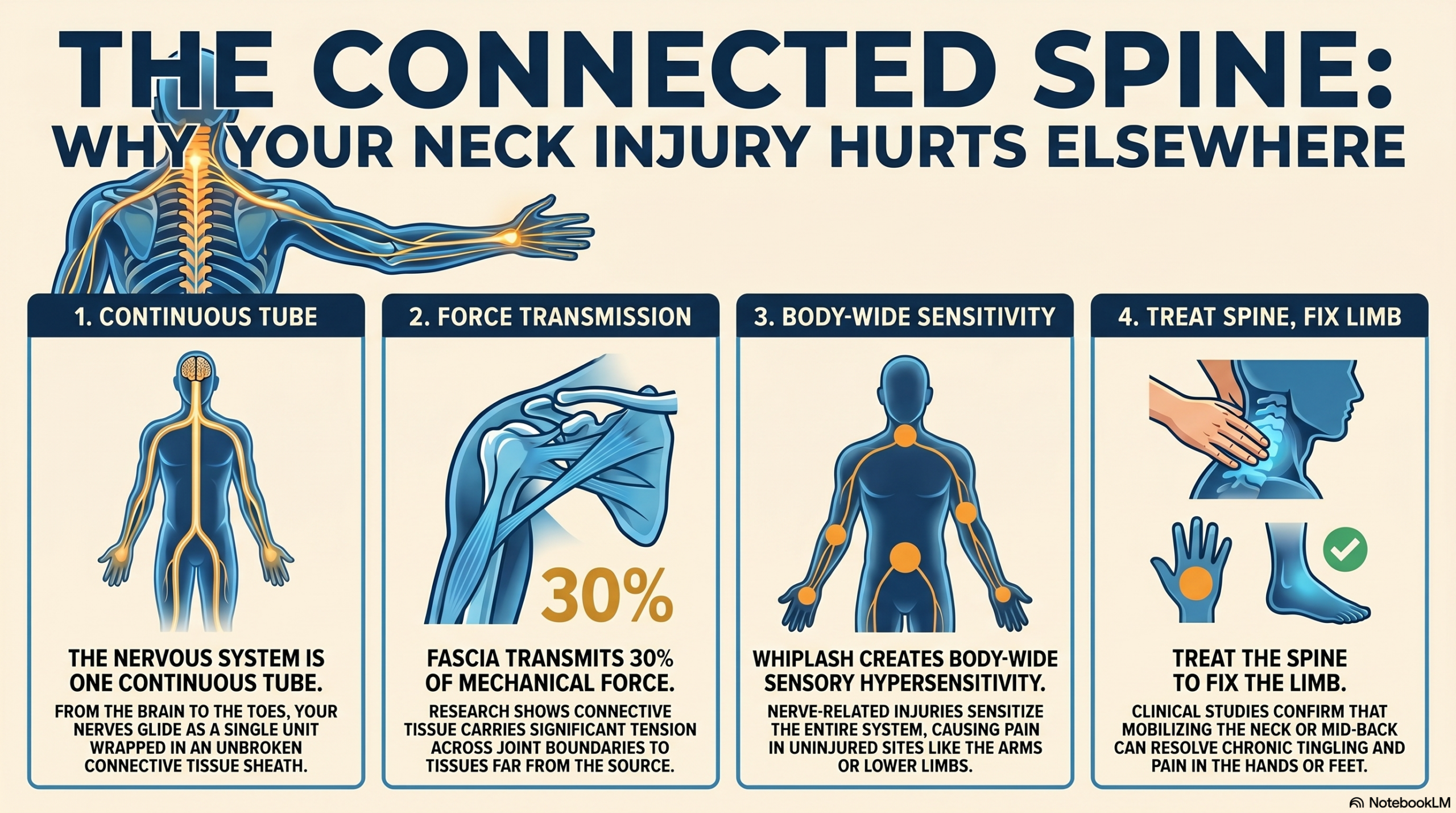
Michele Sterling and colleagues, working out of the University of Queensland, are responsible for some of the foundational work on what they call widespread sensory hypersensitivity after whiplash. In a 2003 study, they showed that people who would go on to recover poorly from whiplash showed measurable sensory abnormalities — increased sensitivity to mechanical pressure and to cold — within the first few weeks of injury. Crucially, the hypersensitivity was not confined to the neck. It showed up over the upper limbs, sometimes the lower limbs, at sites where there had been no direct tissue injury at all.
PMID 12927623 — Sterling M, Jull G, Vicenzino B, Kenardy J. Pain. 2003.
Whiplash patients destined for poor outcome demonstrated generalized sensory hypersensitivity — to pressure and to cold — at remote, uninjured sites within weeks of the original injury.
A follow-up study in 2005 sharpened the finding further. Sterling’s group compared chronic whiplash patients to people with chronic non-traumatic neck pain. Both groups had neck pain, but only the whiplash group showed the widespread, generalized hypersensitivity. The pattern was specific to the way whiplash injures the nervous system, not just the result of any chronic neck pain.
PMID 15722811 — Sterling M, Treleaven J, Edwards S, Jull G. Clin J Pain. 2005.
The generalized hypersensitivity pattern is a fingerprint of whiplash specifically, distinguishing it from other forms of chronic neck pain. The remote sensory changes are tied to the way whiplash injures the nervous system, not to the presence of pain in the neck itself.
The clinical translation is straightforward: when your neck is injured in a way that involves nerve tissue — and almost every whiplash injury involves nerve tissue at some level — the sensitization can show up far from the neck, at sites the original injury never touched. A patient who insists “my neck is fine, it’s my arm” or “my neck is fine, it’s my foot” may be entirely correct about the location of their symptoms and entirely wrong about the location of the underlying problem.
2. The myofascial system is one continuous sheet, not a collection of separate parts
If you have ever dissected a body — most people haven’t, and for understandable reasons — the single most striking visual is that the fascia does not stop. There is no edge. The connective tissue of the neck runs into the connective tissue of the shoulder, which runs into the connective tissue of the back, which runs into the connective tissue of the pelvis, which runs into the connective tissue of the leg, which runs into the connective tissue of the foot, all without a single anatomical break.
A 2019 systematic review by Wilke and colleagues looked specifically at the literature on myofascial chains in the upper limb. They identified three distinct chains running between the neck/shoulder region and the hand, with good anatomical evidence for direct serial tissue continuity all the way through. The implication: tension applied at one end of a chain can be transmitted, mechanically, to the other end of the chain — and an irritation anywhere along the chain can affect tissue tone and movement at a distance.
PMID 31226229 — Wilke J, Krause F, Vogt L, Banzer W. Clin Anat. 2019.
Three distinct fascial chains with confirmed serial continuity were documented between the cervico-shoulder region and the hand, providing an anatomical substrate for force transmission across joint boundaries.
A separate 2016 review by Wilke’s group, looking at intermuscular force transmission along myofascial chains in cadaver and in vivo experiments, estimated that fascia can transmit up to thirty percent of the mechanical force generated by a muscle to tissues at a distance from the muscle itself. That is not a rounding error. That is a meaningful fraction of the work the body does, traveling through structures that anatomy textbooks treat as separate.
PMID 27001027 — Krause F, Wilke J, Vogt L, Banzer W. J Anat. 2016.
Mechanical force generated by one muscle is transmitted through fascial continuity to tissues at a distance, with up to approximately 30% of force traveling beyond the originating muscle’s anatomical insertion.
Frank Willard’s landmark 2012 anatomical review of the thoracolumbar fascia deepens this picture by showing how the lumbar fascia continues, layer by layer, into the paraspinal fascia of the thoracic and cervical regions, eventually fusing with the deep cervical fascia and attaching to the base of the skull. The lumbar back and the upper neck are, fascially speaking, the same continuous sheet. Tension anywhere on the sheet pulls on everywhere else.
PMID 22630613 — Willard FH, Vleeming A, Schuenke MD, Danneels L, Schleip R. J Anat. 2012.
The thoracolumbar fascia is continuous with the paraspinal fascia in the thoracic and cervical regions and fuses with the deep cervical fascia at the cranial base — establishing a single unbroken fascial sheet from sacrum to skull.

3. Mobilizing the thoracic spine improves symptoms in the neck — and in the arm
If the body is one continuous mechanical and neural system, you would expect that working on one region would produce effects in another region. That is, in fact, exactly what the manual therapy literature shows, and it shows it consistently enough that I would call it boring.
A 2014 meta-analysis by Coronado and colleagues — pooling data across multiple randomized trials — found that manual therapy applied to the thoracic spine produced measurable improvements in peripheral outcomes: reductions in arm pain, improvements in shoulder range of motion, and changes in autonomic nervous system markers (skin conductance, blood pressure, heart rate variability) at sites well outside the area being treated. The treatment was local. The effect was systemic.
PMID 25395830 — Coronado RA, Gay CW, Bialosky JE, Carnaby GD, Bishop MD, George SZ. J Electromyogr Kinesiol. 2014.
Cervical and thoracic spinal manual therapy produced measurable changes in pain sensitivity and autonomic markers at sites distant from the manipulated segment — a hypoalgesic effect that extended beyond the treated region.
A 2024 systematic review and meta-analysis by Coppieters and colleagues took this further by comparing manipulation of the cervical spine to manipulation of the thoracic spine for the treatment of neck pain. The somewhat surprising finding: the two approaches produced largely equivalent outcomes. Patients with neck pain improved as much when their thoracic spine was treated as when their neck itself was treated. The implication is not that the neck doesn’t matter — it does — but that the spinal column functions as an integrated system, and that intervening at a distance from the site of pain can produce as much benefit as intervening at the site of pain itself.
PMID 38492291 — Coppieters MW, Hidalgo B, et al. Musculoskelet Sci Pract. 2024.
Across pooled randomized trials, thoracic and cervicothoracic manipulation produced equivalent reductions in pain intensity, disability, and cervical range of motion compared with direct cervical manipulation — confirming that the spine responds as one integrated mechanical system.
4. Neural mobilization works for symptoms at the far end of the cable
I cited the 2017 Basson meta-analysis earlier. There is now a substantial follow-up literature that gets even more specific. A 2025 network meta-analysis by Carmona-Pérez and colleagues evaluated articular plus neural mobilization for cervical radicular pain — the kind of pain that travels down the arm because the nerve root is irritated where it exits the neck. They found that the combination of joint mobilization and neural mobilization, layered onto usual care, produced the largest short-term reduction in pain intensity and disability of any intervention studied.
PMID 40521428 — Carmona-Pérez C, Pérez-Mármol JM, et al. J Orthop Sports Phys Ther. 2025.
Combining articular and neural mobilization with usual care produced the most effective short-term reduction in arm pain and disability across a network of 19 randomized controlled trials of cervical radicular pain.
For the lower extremity, the picture is similar. A 2023 systematic review of neural mobilization in low back and radicular pain — Peacock and colleagues — found small to moderate effect sizes for pain, disability, and straight-leg-raise range of motion when neural mobilization was added to standard care. Treating the nerve, in other words, mattered for symptoms in tissues the nerve supplies.
PMID 36677773 — Peacock M, Douglas S, Nair P. J Man Manip Ther. 2023.
Across thirteen RCTs, addition of neural mobilization to usual care produced significant short-term improvements in pain and disability in low back and radicular pain populations, with effects measurable in distal symptoms including the leg.
5. The slump test reliably identifies a neural component to distal symptoms
I want to come back to that exam-table moment with Maria, because there is a substantial clinical literature behind it. The slump test — flex the spine, flex the neck, extend the knee, dorsiflex the foot, watch what reproduces and what relieves the symptom — is one of the better-studied provocation tests in clinical neurodynamics.
A 2015 diagnostic accuracy study by Urban and MacNeil looked at the slump test’s ability to identify neuropathic involvement in lower-limb pain. Sensitivity was high — around 0.84 — meaning the test rarely misses a true neural component when one is present. The test’s value, in other words, is in confirming that a distal symptom has a neural origin and in pointing back toward the spine as the place that needs treating.
PMID 26221691 — Urban LM, MacNeil BJ. J Orthop Sports Phys Ther. 2015.
The slump test demonstrated sensitivity of 0.84 for identifying a neural contribution to leg pain — a level of accuracy adequate to confirm neural involvement at the bedside when imaging is unavailable or non-diagnostic.
The implication for someone like Maria is that the test bridges the gap that the imaging literature can’t. A standard MRI of her lower back was normal. A nerve conduction study at her ankle was normal. But a thirty-second neurodynamic provocation, done by a clinician who knew what to look for, reproduced her exact foot symptom and pointed up the cable to her neck. That is the test that should have been done in her first week of complaining about her foot. It was not.
The Deeper Science: Why the Body Doesn’t Respect the Anatomy Textbook
To understand why “distance” turns out to be such a poor descriptor of where pain comes from, it helps to think for a moment about what the nervous system actually is, physically.
Picture the human nervous system stripped of everything else — no skin, no muscle, no bone. What you would have is something like a tree. A trunk in the center, where the brain and spinal cord live. Branches radiating outward into every limb, every organ, every patch of skin. The entire structure is bathed in cerebrospinal fluid, surrounded by three protective membranes called the meninges, and tethered to the bony spine at every level by fibrous attachments called denticulate ligaments and dural sleeves.
That central trunk has a startling property: it has to be able to move. When you flex your neck forward, your spinal cord lengthens by several millimeters. When you straighten your leg in front of you, the nerves traveling down to your foot are placed under tension. When you bend forward to tie your shoe, the entire neural tube, from the inside of your skull to the soles of your feet, has to slide and stretch — as one continuous tube — to accommodate the new shape of your spine. The nervous system is not pinned in place. It is engineered to glide.
When that gliding is disrupted — and whiplash is one of the most efficient ways in modern life to disrupt it — the consequences travel along the cable. A small region of inflammation around a cervical nerve root sets up adhesions in the dural sleeve. The dural sleeve loses its glide. Now, every time you flex your neck, you are pulling on a piece of the cable that doesn’t slide the way it used to. The mechanical strain has to go somewhere, and where it goes is downstream — to the next region of the cable that can absorb it, often into the upper limb in the form of arm pain, sometimes into the trunk in the form of mid-back tightness, occasionally — as in Maria’s case — all the way into the leg.
The fascial system reinforces this transmission. The outer covering of the nervous tissue, the epineurium, is not separate from the rest of the body’s connective tissue — it is woven into it. The sleeve around your cervical nerve root is continuous with the sleeve around your brachial plexus, which is continuous with the connective tissue of your shoulder, which is continuous with the deep fascia of your arm, which is continuous with the fibrous envelope of every muscle in your hand. There is no edge. There is no place where the system ends and starts.
This is why strain applied to the lumbar fascia in a cadaver pulls measurably on the cervical fascia at the other end of the spine. It is why a chiropractor can mobilize a patient’s mid-back and watch their shoulder pain change in real time. It is why Maria’s neck injury can broadcast, through a continuous medium, all the way to a small patch of skin on the side of her foot.
The nervous system is one organ shaped like a tree, wrapped in a single sheet of connective tissue that runs from your scalp to your toes. Distance, in this organ, is not what distance is in a road atlas. It is a coordinate inside a single object.

What This Looks Like in Clinic
When a patient like Maria walks into our Petaluma office with a distal symptom that no specialist can explain, the workup is built around the assumption that what looks like a local problem may be a system problem in disguise. Here is what an evaluation actually involves.
Step one: a full history that does not respect the body’s regions. I ask about every injury, every accident, every fall, every surgery for the last decade. Especially I ask about motor vehicle collisions, including the small ones that “didn’t really do anything.” Most patients have forgotten about the fender-bender five years ago. The body has not.
Step two: neurodynamic testing. Slump test for the lower body, upper limb tension tests for the arms, palpation along the brachial plexus, and a careful examination of how the patient’s symptoms change when I add cervical flexion or extension to whatever movement provokes them. If a foot symptom changes when I move the head, that tells me something the foot specialist will never find.
Step three: cervical-thoracic mechanical assessment. I check segmental mobility through the cervical and upper thoracic spine, the cervicothoracic junction, the first and second ribs. I look for the regions of stiffness that have been quietly biasing the neural system for months or years.
Step four: fascial palpation along the relevant chain. If the symptom is in the arm, I trace the dorsal arm chain from the trapezius and infraspinatus through the triceps to the extensor compartment of the forearm. If the symptom is in the leg, I trace the superficial back line from the suboccipital region down to the heel. The hands are looking for restriction, density, and lack of glide — the textures that mark a fascial system that has been under chronic tension.
Step five: foot and pelvis mechanics, if relevant. I look at how the foot pronates, how the pelvis orients during single-leg stance, how the head translates over the shoulders during gait. The thesis stated at the beginning of this article — that treating whiplash sometimes involves the thoracic spine or the foot — is concrete here. If the foot is not absorbing ground reaction force properly, the entire postural chain compensates upward, and the cervical spine is the last segment to pay the bill.
Step six: a treatment plan that addresses the chain, not just the loudest symptom. For Maria, the plan involved cervical-thoracic mobilization, sliders and tensioners for the lumbar dural sleeve, soft-tissue work along her right paraspinal chain, and — yes — a re-evaluation of how her right foot was loading the ground when she ran. Her foot got better as a consequence of treating the rest of her body. Her foot was the symptom. Her foot was not the problem.
A typical course of care for someone like Maria runs eight to twelve visits. The first three visits are usually about confirming the pattern and getting the most aggravated tissues to settle. The middle visits are about restoring glide and motor control across the affected chain. The final visits are about teaching the patient to maintain what we have built, because the chain that learned to broadcast a foot symptom from a neck injury is going to remember how to do that, and the patient’s home program has to be specifically designed to prevent re-running the old pattern.
The Takeaway
If you are reading this and you are carrying around a peripheral symptom that no one can explain — an arm that aches but has a clean MRI, a foot that tingles but has a normal nerve conduction study, a hip that hurts but a clean labrum and a clean gluteus — and somewhere in your history is a motor vehicle collision, even a small one, even one you have stopped thinking about, here is what I want you to consider.
The fact that the symptom is far from the neck does not mean the neck is not involved. The fact that imaging of the symptomatic region is clean does not mean the symptom is imaginary. The fact that three specialists could not find the source does not mean there is no source. It means they were each looking at the wrong end of the cable.
The nervous system is one continuous organ. The fascial system is one continuous sheet. Whiplash is one of the most efficient ways to disrupt both. And the disruption does not stay where it was caused. It travels — sometimes immediately, sometimes over the course of months or years — to the place along the chain that is most able to receive it. That place is rarely where the doctor expects to look.
What to Do Now
If you have been in a motor vehicle collision in the last few years and you have an unexplained peripheral symptom — arm, hand, hip, foot, anywhere — book a movement and neurodynamic evaluation. Not because every symptom traces back to a crash, but because if yours does, none of the specialists you have already seen will have looked for it. In our Petaluma clinic this kind of work is routine, every day.
The body that taught itself to broadcast pain to a distant site can be taught, with patience and the right hands, to stop. The neural cable can be re-mobilized. The fascial sheet can be re-glided. The motor strategy can be re-organized. None of this happens fast, and none of it happens passively, but all of it happens — reliably — when the right region of the body is finally addressed.
Maria came back to clinic eleven weeks after she first walked in. She told me her foot had been quiet for three weeks. She had been running again. She had also, almost as an aside, mentioned that the tight band she had carried across her right shoulder for two years was gone. She had not realized she had a tight band across her right shoulder. She had only realized it was gone when it disappeared.
That is the most common version of this story. People stop noticing chronic tension somewhere along their body’s chain long before they recognize it as part of their original injury. The treatment that fixes the distant symptom often clears, as a byproduct, two or three other low-grade complaints the patient had stopped bothering to mention. None of them were random. All of them were, in their quiet way, the same problem, expressed at different stations along a single continuous wire.
You do not have to live with symptoms whose origin nobody can find. Distance, in the body, is an illusion. The pain in your foot is closer to your neck than your anatomy textbook is willing to admit.
References
- Basson A, Olivier B, Ellis R, Coppieters M, Stewart A, Mudzi W. The Effectiveness of Neural Mobilization for Neuromusculoskeletal Conditions: A Systematic Review and Meta-analysis. J Orthop Sports Phys Ther. 2017. PMID 28704626.
- Sterling M, Jull G, Vicenzino B, Kenardy J. Sensory hypersensitivity occurs soon after whiplash injury and is associated with poor recovery. Pain. 2003. PMID 12927623.
- Sterling M, Treleaven J, Edwards S, Jull G. Widespread sensory hypersensitivity is a feature of chronic whiplash-associated disorder but not chronic idiopathic neck pain. Clin J Pain. 2005. PMID 15722811.
- Wilke J, Krause F, Vogt L, Banzer W. Myofascial chains of the upper limb: A systematic review of anatomical studies. Clin Anat. 2019. PMID 31226229.
- Krause F, Wilke J, Vogt L, Banzer W. Intermuscular force transmission along myofascial chains: a systematic review. J Anat. 2016. PMID 27001027.
- Willard FH, Vleeming A, Schuenke MD, Danneels L, Schleip R. The thoracolumbar fascia: anatomy, function and clinical considerations. J Anat. 2012. PMID 22630613.
- Coronado RA, Gay CW, Bialosky JE, Carnaby GD, Bishop MD, George SZ. Changes in pain sensitivity following spinal manipulation: a systematic review and meta-analysis. J Electromyogr Kinesiol. 2014. PMID 25395830.
- Coppieters MW, Hidalgo B, et al. Cervical manipulation versus thoracic or cervicothoracic manipulations for the management of neck pain: a systematic review and meta-analysis. Musculoskelet Sci Pract. 2024. PMID 38492291.
- Carmona-Pérez C, Pérez-Mármol JM, et al. Effectiveness of Articular and Neural Mobilization for Managing Cervical Radicular Pain: A Systematic Review With Network Meta-Analysis. J Orthop Sports Phys Ther. 2025. PMID 40521428.
- Peacock M, Douglas S, Nair P. Neural mobilization in low back and radicular pain: a systematic review. J Man Manip Ther. 2023. PMID 36677773.
- Urban LM, MacNeil BJ. Diagnostic Accuracy of the Slump Test for Identifying Neuropathic Pain in the Lower Limb. J Orthop Sports Phys Ther. 2015. PMID 26221691.