Movement Before Meaning: Why Restoring Sensation Comes First in Trauma Recovery
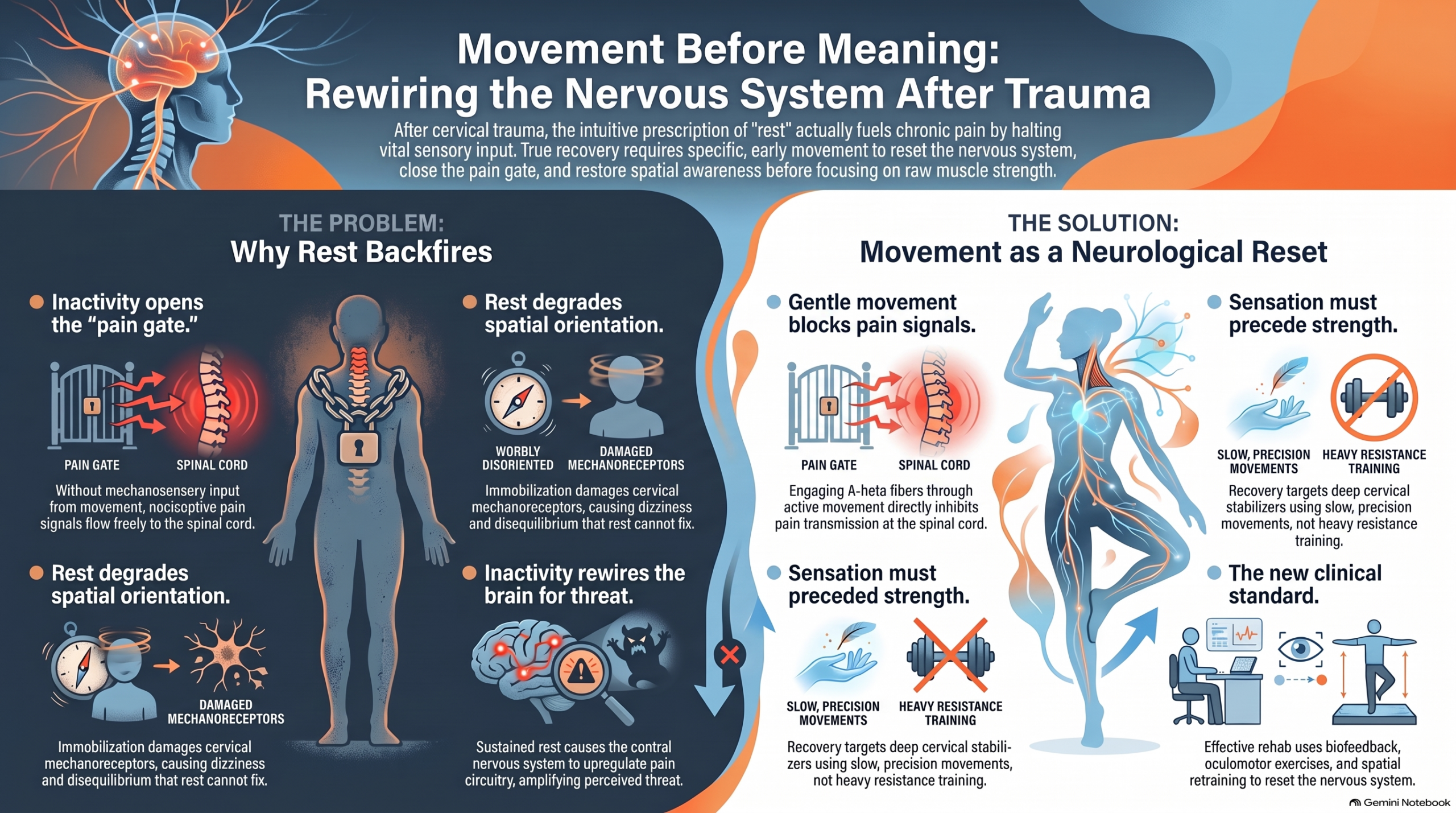
The muscles that protect your spine during a collision are the same muscles that hold you in pain afterward. That is not a paradox — it is physiology. When the cervical paraspinal muscles contract reflexively in the milliseconds following impact, they are doing their job: guarding the spinal cord, limiting displacement, preventing catastrophic injury. But that same protective contraction, sustained past its acute purpose, becomes the architecture of chronic pain. The guarding that saved you is the guarding that keeps you from recovering.
This is the central problem in post-trauma rehabilitation that neither rest nor time resolves: the nervous system, once placed in a protective configuration, does not automatically reset to its pre-injury baseline. It requires a specific input to do so. That input is movement — not aggressive, not heavy, not strength-focused. Just movement. And the evidence for why movement comes first — before meaning, before explanation, before the patient has fully processed what happened — is among the most clinically actionable bodies of research in musculoskeletal medicine.
How Pain Works: The Gate That Movement Controls
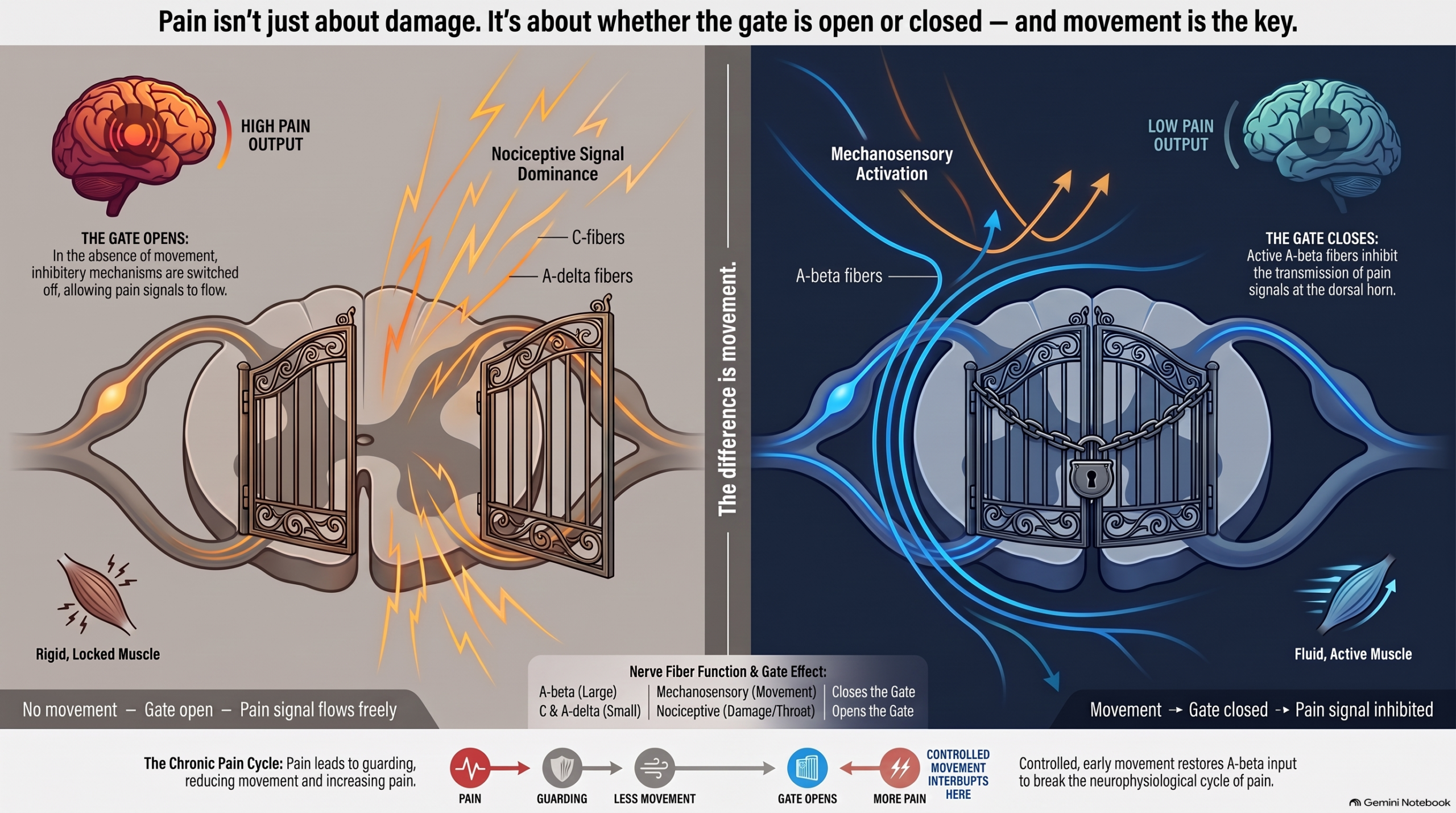
The gate control theory of pain, introduced by Melzack and Wall in 1965, proposed that pain is not a simple signal transmitted from damaged tissue to the brain. It is a regulated output, modulated at the level of the spinal cord by a dynamic interplay between different types of nerve fibers. Large-diameter myelinated fibers — A-beta fibers — carry mechanosensory information: touch, pressure, vibration, movement. Small-diameter unmyelinated or lightly myelinated fibers — C fibers and A-delta fibers — carry nociceptive information: tissue damage signals, inflammatory mediators, threat.
The key insight is this: when A-beta fibers are active, they inhibit the transmission of C-fiber and A-delta signals at the dorsal horn of the spinal cord. The gate closes. When A-beta input is absent — when the region is immobile, guarded, and proprioceptively silent — the gate opens. Nociceptive signals flow more freely. Pain intensity increases not because more tissue is being damaged, but because the inhibitory mechanism has been switched off by the absence of movement.
In the aftermath of cervical trauma, the typical pattern is exactly what the gate control theory predicts would increase pain: the muscles stiffen, movement is restricted, proprioceptive input drops, and A-beta fiber activity decreases. The patient experiences heightened pain — which reinforces the belief that movement is dangerous — which leads to more guarding — which further reduces A-beta input — which opens the gate further. This cycle is not imagined. It is neurophysiological. And it is interrupted by movement.
The clinical implication is direct: the clinician who prescribes rest for a post-collision patient with soft tissue injury is prescribing the conditions that deepen pain. The appropriate prescription — supported by the neurophysiology of pain transmission — is early, controlled, guided movement that restores A-beta fiber input and begins to close the gate.
“The guarding that saved you is the guarding that keeps you from recovering.”
Proprioception: The Hidden Casualty of Cervical Trauma
Proprioception — the body’s internal sense of its own position, movement, and force — is generated primarily by mechanoreceptors embedded in muscle spindles, Golgi tendon organs, joint capsules, and ligamentous tissue. In the cervical spine, these receptors are unusually dense. The suboccipital muscles, which control the relationship between the skull and the upper cervical vertebrae, contain among the highest density of muscle spindles per gram of tissue in the human body. They function less as prime movers and more as precision sensory organs — continuously reporting the position of the head relative to the spine and the spine relative to the trunk.
When cervical soft tissue is traumatized in a rear-end collision, these receptors are directly affected. Capsular disruption at the facet joints alters the mechanical environment of the capsular receptors. Muscle microtears disturb the architecture of the muscle spindle. Edema and inflammation change the chemical environment in which these receptors operate. The result is proprioceptive deficit — a measurable degradation in the accuracy of the cervical spine’s position sense.
Research by Treleaven and colleagues has extensively documented this deficit. Using the joint position error test — in which the subject wears a laser pointer on the head, moves away from neutral, and attempts to return to the starting position with eyes closed — patients with whiplash-associated disorders show significantly greater repositioning error than healthy controls. This is not subjective. It is a measurable spatial error, typically expressed in degrees, that reflects the degraded output of the cervical proprioceptive system.
The consequences of this degradation extend beyond the neck. Because the cervical spine contributes to postural control, gaze stabilization, and the sense of spatial orientation, proprioceptive deficits at this level produce dizziness, visual disturbance, and a diffuse sense of disequilibrium that patients frequently describe and clinicians frequently dismiss as vague or psychosomatic. They are neither. They are predictable downstream consequences of disrupted cervical mechanoreception.
And they do not resolve with rest. They resolve — when they resolve — with specific sensorimotor training that challenges and retrains the position-sense system. Rest does not exercise the system. It allows it to deteriorate further.
Why Rest After Trauma Can Backfire
The intuitive prescription for pain is rest. Pain signals danger; danger signals the need to stop. This logic is sound for acute injury in which continued loading would extend tissue damage — a fresh fracture, an acute herniation compressing a nerve root, a torn tendon in the hours following injury. In these cases, protection and offloading are genuinely therapeutic.
It is not sound for soft tissue cervical injury after the acute phase has passed.
The evidence on this point is consistent across multiple randomized controlled trials. Studies comparing cervical collars to early active mobilization show significantly worse outcomes in the collar group — in terms of both symptom duration and functional recovery — despite the fact that wearing a collar feels, to the patient, like appropriate protection. Schnabel and colleagues, in a randomized controlled outcome study, found that patients assigned to active mobilization had significantly better outcomes on pain, range of motion, and disability scores at six weeks compared to collar therapy. The collar provided comfort. It did not provide recovery.
The mechanism is now well understood. Immobilization produces rapid changes in connective tissue: collagen fibers become disorganized, cross-link in dysfunctional patterns, and lose the structural alignment that allows ligamentous tissue to sustain load. Muscle fibers undergo atrophy and fatty infiltration — changes that, in the cervical multifidus, have been documented in whiplash patients using fat-fraction MRI analysis by Elliott and colleagues. The deep cervical stabilizers that control segmental motion reduce their activation, creating proprioceptive gaps and mechanical instability that sustain the pain cycle.
Beyond the structural changes, immobility has direct neurological effects. Central sensitization — the upregulation of the central nervous system’s pain-processing circuitry — develops more readily in the context of sustained inactivity and proprioceptive deprivation. The dorsal horn neurons that process nociceptive input from the cervical spine become more excitable. Inhibitory pathways become less active. The brain’s threat map assigns greater danger value to sensory input from the neck — which means that the same stimulus perceived as mild pressure in a healthy, active individual is perceived as intense pain in a patient whose nervous system has been reorganized by weeks of protective rest.
This is what it means to say that rehabilitation must happen before the patient fully cognitively processes the trauma. The window in which movement can interrupt central sensitization before it becomes consolidated is not indefinite. The earlier sensorimotor input is restored, the less reorganization the nervous system undergoes. Waiting for pain to subside before beginning movement reverses the causal sequence: movement is what causes pain to subside.
Movement as a Neurological Reset
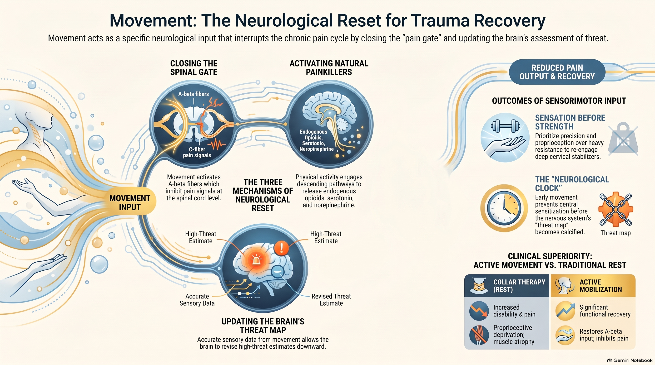
The therapeutic mechanism of movement in post-trauma rehabilitation is not primarily mechanical — it is neurological. Controlled movement does not simply stretch tight tissue or pump edema out of a joint. It generates the specific type of afferent input that the nervous system requires to recalibrate its threat output.
This recalibration operates through several concurrent mechanisms.
First, movement activates the large-diameter A-beta fibers that carry mechanosensory information, directly engaging the gate-closing mechanism at the dorsal horn. Active cervical movement — even within a reduced range — generates proprioceptive signals that compete with and inhibit nociceptive transmission. The relief patients report during gentle manual therapy or controlled cervical mobilization is not placebo; it reflects actual gate-level inhibition of pain transmission.
Second, movement activates the descending pain modulation system — the network of pathways originating in the periaqueductal gray matter and rostral ventromedial medulla that inhibit nociceptive processing at the level of the spinal cord. This system uses endogenous opioids, norepinephrine, and serotonin as its primary neurotransmitters. Conditioned pain modulation studies have demonstrated that this system is compromised in patients with chronic whiplash-associated disorders — and that its responsiveness can be partially restored through sensorimotor training.
Third, movement provides the brain with updated information about the state of the body. The predictive processing framework of pain proposes that pain is the output of the brain’s threat assessment — not the input from damaged tissue. When the brain lacks accurate information about the body — when proprioceptive signals are absent, distorted, or inconsistent — it defaults to a higher threat estimate, which is expressed as pain. Movement corrects the information. It updates the brain’s model of the body. It provides evidence that the body is capable of function, which the brain uses to revise its threat estimate downward.
This is the neurological argument for why movement must come before meaning. Before the patient understands what happened — before the cognitive and emotional processing of the trauma is complete — the nervous system can be influenced by sensorimotor input. The window for this influence is early. It is concrete. And it does not wait for psychological readiness.
What Recovery Actually Looks Like: Sensation Before Strength
One of the most consequential clinical errors in post-trauma rehabilitation is organizing recovery around strength as the primary outcome. Strength-focused rehabilitation — resistance exercises, progressive loading, return-to-activity benchmarks — is appropriate for contexts where the primary deficit is tissue weakness. It is not the appropriate framework for cervical spine rehabilitation following whiplash injury, where the primary deficits are proprioceptive and sensorimotor.
Research by Jull and colleagues at the University of Queensland established that patients with chronic whiplash-associated disorders show specific impairment in the activation of the deep cervical flexors — the longus colli and longus capitis — relative to the superficial cervical flexors. This impairment is not simply weakness in the conventional sense. It reflects a disruption in motor control pattern: the deep stabilizers, which should be the first to activate and the last to relax, have reduced their activity in response to the sensory disruption caused by the injury. The superficial muscles compensate. This compensation is mechanically inefficient, proprioceptively impoverished, and energetically costly — and it perpetuates the pain cycle by loading structures that are already sensitized.
The therapeutic target is not to strengthen the superficial muscles — they are already overactive. The target is to retrain the deep cervical stabilizers. This is accomplished not through resistance but through precision: slow, controlled, proprioceptively demanding movements that force the deep system to activate, sustain, and coordinate its output. Cranio-cervical flexion training — the specific intervention studied by Jull and colleagues — uses a pressure biofeedback unit to provide real-time sensory feedback about the depth and control of the deep flexor activation. It is sensation-guided, proprioception-informed, and produces better outcomes for pain and function than conventional strength training in the post-whiplash population.
Recovery, correctly understood, is the progressive restoration of accurate sensorimotor information — from the injury site to the spinal cord to the brainstem to the cortex. Strength follows from that restoration. It does not precede or substitute for it.
Clinical note: The deep cervical flexors are the primary rehabilitation target after whiplash — not because they are weak, but because the injury disrupts their motor control pattern. Biofeedback-guided cranio-cervical flexion training retrains activation depth and timing, which standard strengthening cannot replicate.
The Clinical Standard: Rehabilitation That Reawakens
The clinical standard for post-trauma cervical rehabilitation must be built around sensorimotor retraining as the primary intervention, not as an adjunct to strength work. In practice, this includes:
Cranio-cervical flexion training — biofeedback-guided retraining of deep cervical flexor activation, with Level 1 evidence for reducing pain and disability in chronic whiplash-associated disorders from the randomized controlled trial by Jull and colleagues (2009).
Joint position error retraining — laser or digital pointer feedback that progressively challenges and improves proprioceptive repositioning accuracy. Treleaven and colleagues demonstrated that this intervention improves repositioning accuracy, reduces dizziness, and decreases pain.
Oculomotor training — exercises targeting the coordination between eye movement and head movement, addressing the cervico-ocular reflex and gaze stabilization impairments responsible for much of the dizziness and visual disturbance in whiplash-associated disorder.
Graded sensorimotor exposure — progressive, controlled exposure to movement in postures and contexts that have become pain-associated, using the principles of graded motor imagery and pain neuroscience education to recalibrate the brain’s threat response without triggering sensitization.
Each of these interventions shares a common architecture: they provide precise sensory input to a system that has been deprived of accurate information, and they measure the output — repositioning accuracy, activation depth, eye movement control — rather than effort alone. They are rehabilitation tools for a nervous system.
This is what distinguishes modern cervical rehabilitation from the rest-and-strengthen model: it targets the actual deficits caused by the actual injury mechanism. And it begins early — before pain fully resolves, before full range of motion is restored, and before the patient’s cognitive map of the injury is complete.
Clinical documentation standard: Record joint position error, cervical range of motion, deep flexor activation level, and pain scores at every post-collision evaluation. These functional measures are the evidentiary record of what imaging cannot capture — and they are the metrics that predict who recovers and who does not.
Conclusion
The phrase “movement before meaning” is not a philosophy — it is a clinical principle grounded in gate control theory, proprioceptive neuroscience, and the evidence base for early active rehabilitation in whiplash-associated disorders. It describes the sequence that the nervous system requires: sensorimotor input first, cognitive processing second.
The patient who rests, waits for understanding, and begins rehabilitation only after the pain makes sense is working against the neurological clock. Central sensitization consolidates in the space that movement could have occupied. Proprioceptive deficits deepen. The brain’s threat map calcifies around the injury.
The patient who moves — carefully, precisely, with clinical guidance — is giving their nervous system the input it needs to revise its output. The gate closes. The descending modulation system activates. The deep cervical stabilizers re-engage. The brain receives evidence that the body can function, and it lowers its alarm.
This is not about pushing through pain recklessly. It is about understanding what pain is — a protective output of the nervous system, not a measure of ongoing damage — and providing the inputs that revise that output downward. Movement is the primary input. Meaning follows. Recovery follows meaning. But recovery cannot begin until movement has already started.
References
- Melzack R, Wall PD. Pain mechanisms: a new theory. Science. 1965;150(3699):971–978.
- Treleaven J, Jull G, Sterling M. Dizziness and unsteadiness following whiplash injury: characteristic features and relationship with cervical joint position error. Journal of Rehabilitation Medicine. 2003;35(1):36–43.
- Jull G, Falla D, Vicenzino B, Hodges PW. The effect of therapeutic exercise on activation of the deep cervical flexor muscles in people with chronic neck pain. Manual Therapy. 2009;14(6):696–701.
- Elliott JM, Pedler AR, Kenardy J, et al. The temporal development of fatty infiltrates in the neck muscles following whiplash injury. PLoS ONE. 2011;6(8):e21194.
- Moseley GL, Butler DS. Fifteen years of explaining pain: the past, present, and future. Journal of Pain. 2015;16(9):807–813.
- Schnabel M, Ferrari R, Vassiliou T, Kaluza G. Randomised, controlled outcome study of active mobilisation compared with collar therapy for whiplash injury. Emergency Medicine Journal. 2004;21(3):306–310.
- Treleaven J. Sensorimotor disturbances in neck disorders affecting postural stability, head and eye movement control. Manual Therapy. 2008;13(1):2–11.