Seatbelt Injuries Beyond the Obvious Bruise
The device that saved your patient’s life also created a predictable constellation of injuries that expire undocumented in two weeks.
The Trade-Off Built Into Every Car
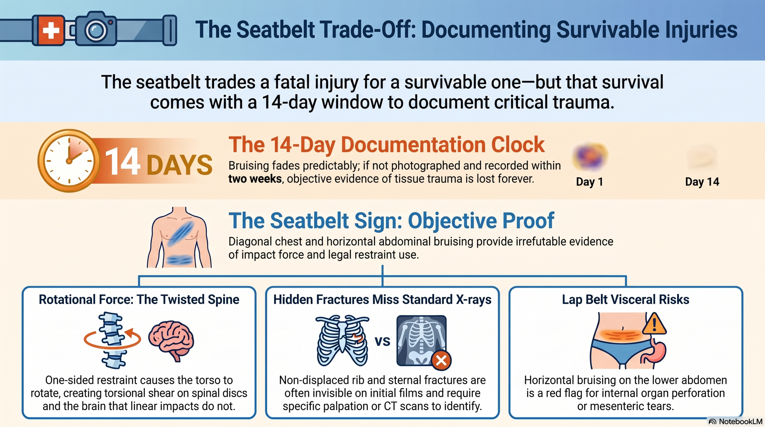
The same device that kept you alive is the reason you have a rib fracture. That is not a side effect of a design flaw — it is the functional trade-off built into every seatbelt in every vehicle produced for the last six decades.
Seatbelts work by arresting momentum. When the vehicle decelerates suddenly, the belt holds the body in place while the collision energy dissipates elsewhere. In the absence of a seatbelt, that energy goes into the windshield, the dashboard, or the road. With a seatbelt, it goes into the tissues the belt contacts directly — the chest wall, the clavicle, the shoulder, the abdomen — and into the rotational forces produced when one shoulder is held fixed while the rest of the body continues moving.
This is not an argument against seatbelts. General Patton died in 1946 in a low-speed urban collision because he was not wearing one. Modern crash biomechanics confirm the same conclusion across millions of data points every year. The seatbelt trades a fatal injury for a survivable one — and that trade is worth making. But preventing death is different from preventing injury. The survivable injuries the seatbelt creates are among the most systematically under-documented findings in post-collision medicine.
This article is about the injuries that seatbelt restraint causes. Not the injuries it prevents — those are well understood. The injuries it causes: the bruising that expires in two weeks and is worth more than a report, the rib fractures that don’t appear on the first X-ray, the rotational spinal forces that are mechanically inevitable and clinically undocumented, and the shoulder pathology that was contacted directly by the belt and never examined. These injuries are predictable. They follow from the mechanism with the same reliability that posterior disc herniations follow from rear-end flexion-extension loading. The question is whether the provider is looking for them.
“Preventing death is different from preventing injury. The seatbelt trades a fatal injury for a survivable one — and the survivable injuries it creates are almost never documented.”
The Seatbelt Sign: Evidence That Has an Expiration Date
The seatbelt sign is the visible marking left when belt contact forces are sufficient to contuse the soft tissues beneath the belt. It appears in two geometrically distinct patterns corresponding to the two belt components.
The shoulder belt bruise runs diagonally across the anterior chest — from the restrained shoulder downward toward the opposite lower rib cage. In a standard US driver-side configuration, this runs from the left shoulder toward the right lower ribs. The pattern is pathognomonic: its shape cannot be produced by a fall, a punch, or a sports injury. It matches the exact geometry of the shoulder belt. When it is present, it communicates three simultaneous facts: the patient was properly restrained, the forces were sufficient to contuse soft tissue through clothing, and the structures directly beneath the belt contact zone are at risk for deeper injury.
The lap belt bruise runs horizontally across the lower abdomen or pelvis, corresponding to the lap portion of the belt. This pattern carries a more serious clinical implication: the abdominal viscera sit immediately beneath it, protected only by the abdominal wall musculature. Lap belt bruising should immediately trigger visceral screening.
Documentation window: Bruising evolves predictably — red at 24 hours, purple by day 3, green by day 7, yellow by day 10, gone by day 14. After it fades, it is gone permanently unless it was photographed. Most patients don’t present within 48 hours. Inspect for seatbelt bruising at every post-collision intake, photograph it if present, and document its verbal history if it has already resolved.
Insurance valuation systems — including Colossus, the algorithm widely used by major carriers — weight objective, visible injury evidence more heavily than subjective symptom reports. A photograph of seatbelt bruising is objective evidence of tissue trauma. It documents injury at the moment it is visible. It is not a symptom report that can be disputed — it is a visual record that establishes force magnitude, mechanism, and contact location simultaneously.
The photography protocol is straightforward: obtain consent, photograph in the clinic, include a dated marker or identifying element in at least one image, take multiple angles (one contextual showing anatomical location, one close-up showing color and extent), and document dimensions and color in the written record. If the patient presents after bruising has resolved, the verbal history is the documentation: “Patient reports diagonal ecchymosis across anterior chest consistent with shoulder belt pattern, present for approximately eight days post-collision, now resolved. Evolution consistent with typical ecchymosis resolution timeline.”
The Rotation Mechanism: The Injury the Belt Creates
The rotation mechanism is the most clinically significant and most consistently undocumented consequence of seatbelt restraint in a frontal or near-frontal collision. It is not a complication — it is mechanically inevitable, and it occurs in every restrained occupant whose vehicle decelerates in a direction non-symmetric relative to the belt attachment.
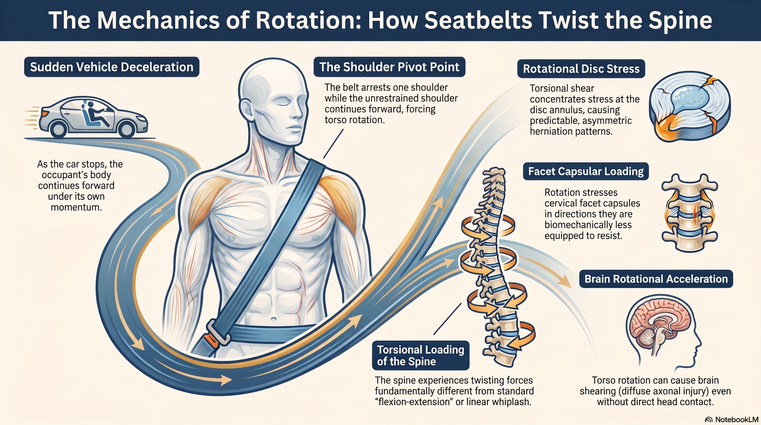
The mechanism: the vehicle decelerates. The shoulder belt arrests the left shoulder. The right shoulder continues forward under its own momentum. The torso rotates around the left shoulder as the pivot point. The cervical and thoracic spine experience torsional loading — forces applied in a twisting direction that differ fundamentally from the flexion-extension forces of standard rear-end whiplash.
The clinical consequences of this rotation are specific:
- Rotational disc stress. Intervertebral discs tolerate axial compression and moderate flexion-extension. They are significantly less tolerant of torsional shear, which concentrates stress at the outer annular fibers. Herniation patterns produced by rotational loading are more likely to be posterolateral or lateral, and asymmetric in the direction of the rotation.
- Facet capsular loading in rotation. Cervical facet capsules are implicated in approximately 55% of chronic neck pain following whiplash injuries (Lord et al., Spine). In a restrained collision with obligate rotation, the facet capsules are additionally stressed in torsion — the direction they are biomechanically least equipped to resist.
- Brain rotational acceleration. Diffuse axonal injury is produced more reliably by rotational acceleration of the brain than by linear acceleration. Restrained occupants who experience torso rotation create conditions for brain rotation even without head contact.
Causation language: “Driver restrained by three-point seatbelt in frontal collision. Restraint of left shoulder with continued forward movement of right shoulder produces obligate clockwise torso rotation, generating torsional loading of the cervical and thoracic spine. Clinical findings of asymmetric cervical rotation restriction are consistent with this mechanism.”
The documentation failure here is institutional. Providers note that the patient was wearing a seatbelt and move on. They do not write the sentence that follows: that seatbelt restraint of the left shoulder in this frontal collision produced obligate clockwise torso rotation, loading the cervical and thoracic facet joints in torsion and concentrating rotational shear forces at the disc annulus in a rightward direction. That sentence is not opinion — it is applied biomechanics. And it is the foundation of the causation argument that ties the mechanism to the imaging findings to the clinical presentation.
Rib and Sternal Injuries: The Fractures That Don’t Appear on First X-Ray
The shoulder belt is two to three inches wide. In a significant deceleration, it applies the momentum of the entire upper body across that narrow strip. The anterior rib cage and clavicle sit directly beneath it. Both are at risk for fracture. Both are routinely missed.
Rib fractures from belt restraint present a specific diagnostic challenge: non-displaced fractures are notoriously difficult to identify on plain chest radiographs. The fracture line may not be visible at all in the first 48 hours, becoming apparent radiographically only as periosteal callus forms 7–10 days after injury. CT is three to five times more sensitive than plain X-ray for non-displaced rib fractures — but CT isn’t ordered unless someone specifically suspects a rib fracture, which requires palpating the ribs, which requires knowing the belt creates rib fractures.
Clinical presentation:
- Sharp, well-localized chest pain that intensifies with deep inspiration or coughing
- Pain with trunk rotation
- Reproducible point tenderness on direct palpation at the fracture site
- Possible palpable crepitus at the fracture site with movement
- Guarded, shallow breathing to avoid the pain
Palpate the anterior ribs in the belt contact zone on every patient with seatbelt sign. Document point tenderness precisely. Order chest X-ray; follow with CT if plain film is non-diagnostic and clinical suspicion remains high.
A documented rib fracture — even non-displaced — is a specific, objective diagnosis. It requires specific management: restricted activity to allow healing, breathing exercises to prevent pneumonia from splinted respiration, and follow-up imaging. It also establishes that the collision forces were sufficient to fracture bone — a finding that has material implications for case valuation and that directly contradicts any defense argument that the crash was minor.
Sternal injuries deserve separate attention. The sternum sits along the belt path at the chest midline. Sternal fractures present with deep, central chest pain; exquisite tenderness on direct palpation; and pain with any shoulder girdle movement. Standard cervical and lumbar X-ray series do not visualize the sternum — a dedicated lateral sternal view or CT is required. Sternal fractures are also associated with myocardial contusion; any patient with sternal tenderness requires physician evaluation before chiropractic management proceeds.
“A documented rib fracture proves the collision forces were sufficient to break bone. A missing rib fracture diagnosis, when the fracture exists and was not imaged, proves nothing — except that the right study was never ordered.”
The Shoulder Complex: Three Structures at the Belt Contact Point
The shoulder belt contacts three anatomically distinct structures at the shoulder: the clavicle, the acromioclavicular joint, and the rotator cuff region. Each has a specific and documented injury pattern tied to the belt contact mechanism. Each is routinely unexamined because clinical attention focuses on the cervical spine.
- Clavicle fractures: The belt applies concentrated restraining force along the clavicular shaft. Mid-shaft clavicle fractures present with focal point tenderness, pain with shoulder movement, and possible step-off deformity. Dedicated clavicle views are required; they are not captured on standard cervical X-ray series.
- Acromioclavicular joint injuries: The belt applies a downward and inward force at the shoulder that replicates the mechanism of AC separation. Examination findings: direct tenderness over the AC joint, pain with horizontal adduction (cross-body reach test), and the piano key sign. A Grade II AC separation is a torn ligament that produces chronic shoulder dysfunction if undiagnosed and untreated.
- Rotator cuff injuries: At the moment of impact, the rotator cuff contracts maximally to stabilize the shoulder against sudden loading — an eccentric contraction under abrupt high load, the mechanism most reliably associated with rotator cuff tears. In patients over 40, pre-existing degenerative thinning lowers the rupture threshold. A negative initial shoulder examination does not rule out rotator cuff pathology; if shoulder symptoms persist, dedicated shoulder MRI is indicated.
- Brachial plexus involvement: The belt can apply traction to the brachial plexus during the restraint event, producing arm weakness, paresthesia, and the “dead arm” presentation. Electrodiagnostic testing is indicated when involvement does not resolve.
Documentation mandate: The mechanism predicts shoulder involvement whenever the shoulder belt contacts the shoulder. The examination must be performed regardless of patient complaint. “Mechanism involves direct belt contact to right shoulder; shoulder examination performed regardless of initial chief complaint” belongs at the top of the shoulder examination section of every seatbelt-sign intake note.
Visceral Trauma: When the Clinical Finding Is a Referral
The lap belt restrains the pelvis and lower abdomen. When the vehicle decelerates, the abdominal viscera continue forward against the belt. In significant decelerations, this produces injuries outside the scope of musculoskeletal management:
- Bowel perforation: Direct belt compression can produce full-thickness perforation of the small bowel, leading to peritoneal contamination — a surgical emergency.
- Mesenteric tears: Tearing produces vascular injury and bowel ischemia.
- Solid organ contusion: Liver and spleen contusion produces hemorrhage that may expand over hours to days.
- Bladder rupture: A full bladder at the time of impact is particularly vulnerable to compression injury.
Red flags requiring immediate emergency department referral: abdominal pain worsening over time, nausea or vomiting, abdominal rigidity or guarding, diffuse tenderness on palpation, any signs of hemodynamic instability. If a patient presents with lap belt bruising and any abdominal complaint, refer before treating. Document the referral explicitly.
Documentation Protocol: The Two-Week Clock
The seatbelt injury pattern creates a documentation opportunity that begins at the moment of collision and closes approximately two weeks later. Most of the time, no one captures it — because no one built a protocol that systematically looks for it.
Five questions at every post-collision intake:
- “Were you wearing your seatbelt?” — Documents compliance; determines which shoulder was restrained; establishes the rotation direction.
- “Do you have any bruising from the seatbelt?” — Inspect regardless of answer; photograph if present; document dimensions, color, and location.
- “Does it hurt to take a deep breath or cough?” — A yes triggers rib palpation, point tenderness documentation, and a chest X-ray order.
- “Do you have any shoulder pain on the side the belt crossed?” — Examine the shoulder regardless of the answer; document AC joint, rotator cuff, and glenohumeral findings specifically.
- “Any abdominal pain or nausea?” — With lap belt bruising, a positive answer requires immediate referral.
The causation documentation that ties everything together: write the belt geometry explicitly. Which shoulder was restrained. Which direction the torso rotated. Which structures were contacted directly. Connect each clinical finding to the mechanism that produced it. “The shoulder belt, crossing from the left shoulder diagonally to the right lower chest, contacted the anterior left 5th through 7th ribs directly. Point tenderness at the anterior 5th rib is consistent with direct belt contusion; chest X-ray ordered to evaluate for fracture. Restraint of the left shoulder with continued forward movement of the right shoulder produced clockwise torso rotation; this mechanism is consistent with the asymmetric cervical range-of-motion restriction documented on examination, with greater restriction in right lateral flexion and left rotation.”
That is a complete causation statement. It links the mechanism to the anatomical contact to the clinical findings. It holds up.
The Legal Calculus: Why Photographs Change Case Value
Insurance valuation systems process inputs and weight objective, documented evidence over subjective complaint. The documentation hierarchy, from highest to lowest impact on case valuation:
- Photographs of visible injury — A photograph of diagonal chest bruising is a visual record of tissue hemorrhage at a specific anatomical location, produced by a specific mechanism. It establishes force magnitude, contact geometry, and injury location. It cannot be disputed the way a pain report can be disputed.
- Specific diagnoses from imaging — A documented rib fracture is categorically different from “chest wall pain.” It proves the forces were sufficient to break bone and commands materially different case valuation.
- Rotation mechanism documentation — When the report documents seatbelt-produced rotation and the MRI shows a disc herniation in the direction predicted by that rotation, the causation chain is complete and defensible.
- Shoulder findings at the belt contact point — An AC joint separation in the shoulder directly contacted by the belt is mechanism-specific, causally irrefutable, and often determinative for case value.
Two patients in identical crashes — one whose provider photographed the bruising, palpated the ribs, imaged the shoulder, and wrote the causation statement; one whose provider wrote “cervical and thoracic pain following motor vehicle collision” — will have materially different case outcomes. Not because their injuries differ. Because their documentation does.
Why This Pattern Is Consistently Missed
The under-documentation of seatbelt injuries follows a predictable pattern:
- Training focus on the cervical spine. Post-collision clinical education emphasizes cervical mechanics and whiplash biomechanics. It does not systematically teach the seatbelt injury pattern. Providers find what they were trained to find.
- Patient self-reporting gaps. Patients in acute cervical pain do not parse their body’s injury signals. The rib tenderness they feel with deep breaths is “just from the accident.” They need to be asked specifically.
- The bruising timeline. Optimal documentation requires catching bruising within the first week. Providers who wait for the patient to mention it will miss the window.
- Imaging protocol defaults. Standard cervical X-ray series does not include the anterior ribs, sternum, or clavicle. Without a specific clinical indication driving a specific order, these structures go unimaged — and undiagnosed fractures are legally indistinguishable from absent ones.
- The ER clearance fallacy. “The ER cleared me” means the patient was not in immediate danger of dying. It does not mean the ribs are intact or the shoulder is uninjured.
Conclusion: The Belt Tells the Story — If Someone Records It
Every patient who was wearing a seatbelt at the time of collision carries a mechanism roadmap. The belt geometry predicts the rotation. The rotation predicts the spinal injury pattern. The contact points predict the rib, sternal, clavicular, and shoulder pathology. The lap belt predicts the visceral risk. The two-week window for visible documentation defines the urgency.
The protocol is not complicated: five questions at intake, visual inspection and photography, rib palpation, shoulder examination, abdominal screening, and a causation statement that connects the belt geometry to each clinical finding. Imaging orders that follow the mechanism, not just the symptom.
The seatbelt saved your patient’s life. It also told the story of their injuries — in bruise geometry, in fracture lines, in rotational spinal loading, and in shoulder pathology at the exact contact point. That story is available for approximately two weeks. After that, it fades permanently.
The providers who find it, capture it, image it, and write it into the record are the ones whose patients have complete cases. The providers who don’t are the ones whose patients settle for muscle strain — when they actually had rib fractures and AC separations and rotational disc injuries that no one ever found.
“The seatbelt saved their life. It also left evidence of exactly what happened to them. That evidence is visible, photographable, and documentable for about two weeks. After that, it fades.”
The question is whether anyone is reading it while the evidence is still visible.
References
- Lord SM, Barnsley L, Wallis BJ, Bogduk N. Chronic cervical zygapophysial joint pain after whiplash. Spine. 1996;21(15):1737–1745.
- Grauer JN, Nightingale RW, Pintar FA, et al. Whiplash produces an S-shaped curvature of the neck with hyperextension at lower levels. Spine. 1997;22(21):2489–2494.
- Gennarelli TA, Thibault LE. Biomechanics of acute subdural hematoma. Journal of Trauma. 1982;22(8):680–686.
- Teasdale G, Jennett B. Assessment of coma and impaired consciousness: a practical scale. Lancet. 1974;2(7872):81–84.