The Hidden Brain Injury: What Your Sense of Smell Reveals After a Car Crash
Cranial Nerve I is the most commonly injured cranial nerve in motor vehicle collisions and the one most consistently omitted from post-collision examinations — despite a thirty-second test that reveals its damage and a direct anatomical connection to the memory problems patients report.
The Most Common Nerve Injury Nobody Tests For
The olfactory nerve is the most commonly injured cranial nerve in motor vehicle collisions. It is also the cranial nerve least likely to appear in any post-collision clinical record.
Those two facts exist simultaneously in clinical practice — not because the injury is subtle or difficult to detect, but because detecting it requires a test that takes less than a minute and almost nobody orders it.
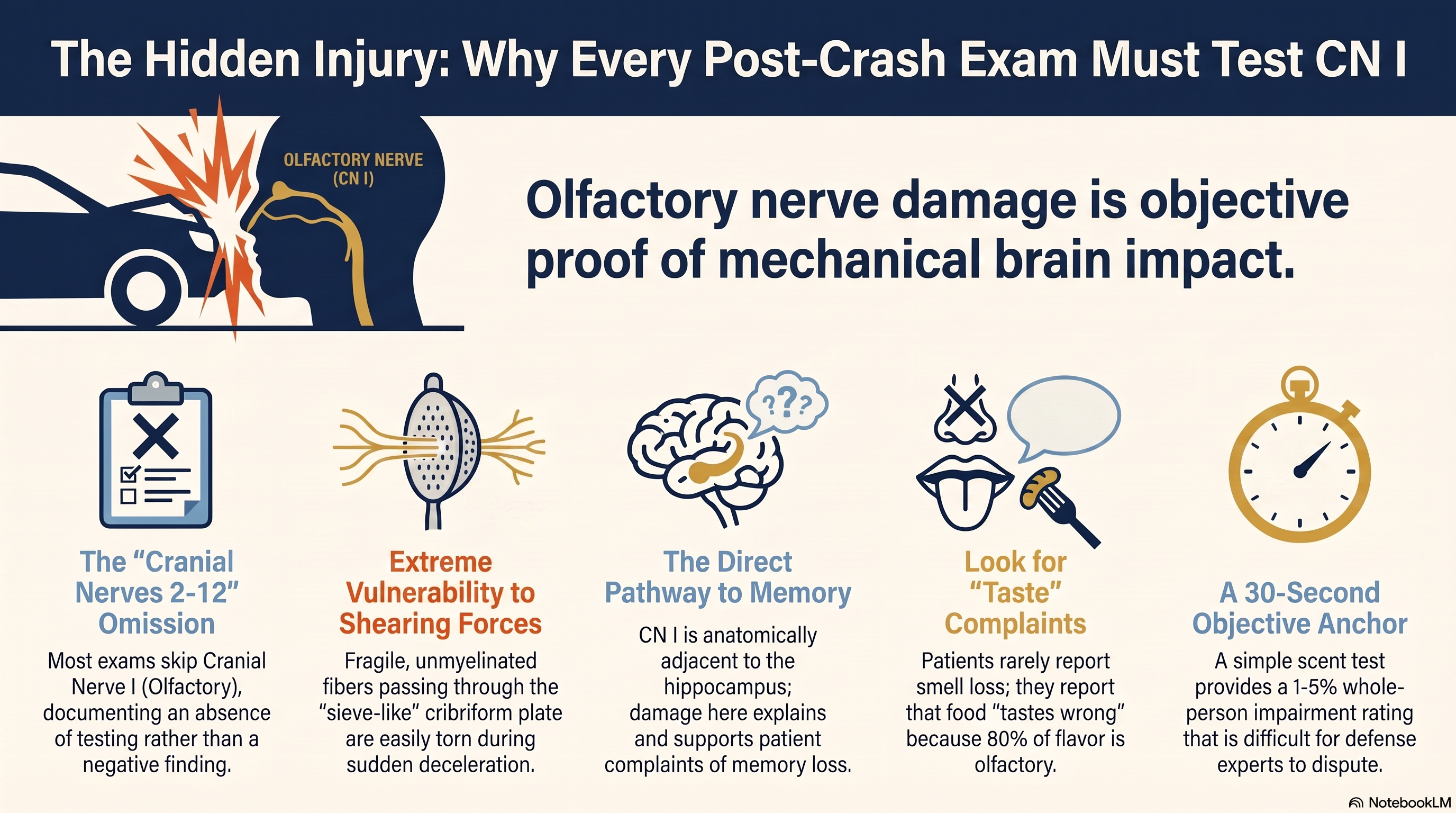
The standard post-collision neurological notation reads: “Cranial nerves 2 through 12 grossly intact.” That sentence is written in tens of thousands of emergency department charts, chiropractic intake forms, and primary care records every year. It sounds complete. It is not. Cranial nerve 1 — the olfactory nerve — is not in that range. It has been omitted from the assessment entirely. The note does not document a negative finding for CN I. It documents the absence of an examination.
This distinction matters clinically, medically, and legally, because olfactory nerve injury following a motor vehicle collision is not a curiosity. It is a predictable, mechanistically sound consequence of the forces that act on the brain during impact — and it is diagnostically connected, through anatomy, to exactly the kinds of cognitive complaints that post-collision patients describe and defense experts dismiss: short-term memory problems, concentration difficulties, and the sense that something in their head has changed since the crash.
The test that identifies olfactory nerve damage takes thirty seconds and requires no equipment that is not already in the room.
Why the Olfactory Nerve Is So Vulnerable
Understanding why Cranial Nerve I is commonly injured requires understanding how it enters the skull — and what happens to it when the brain moves independently of that skull.
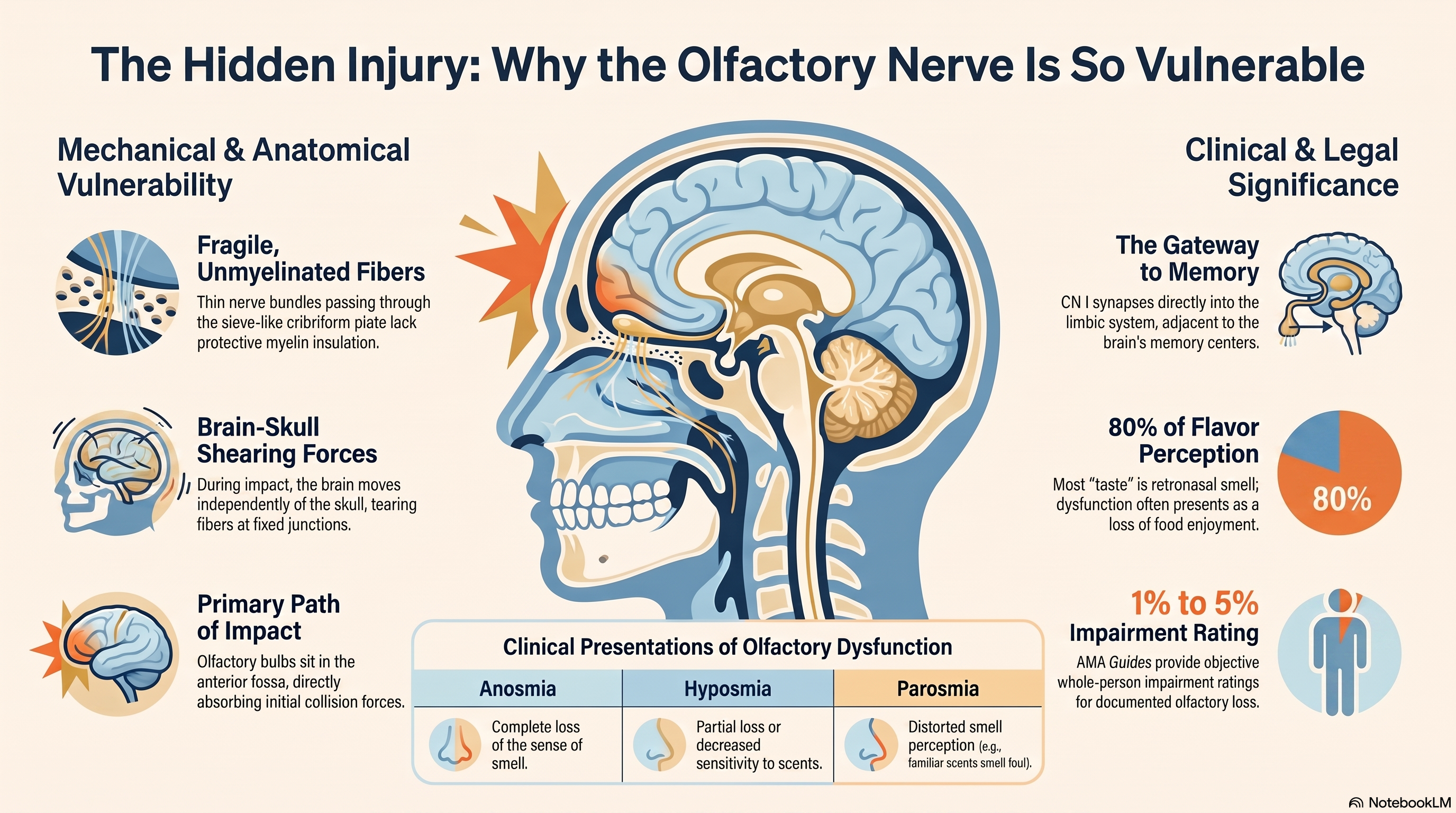
The olfactory nerve is not a single trunk like most cranial nerves. It is a bundle of unmyelinated nerve fibers — the olfactory fila — that originate in the olfactory epithelium of the nasal cavity, pass upward through the perforations of the cribriform plate (a thin, sieve-like section of the ethmoid bone at the base of the skull), and synapse with the olfactory bulb, which sits directly on the floor of the anterior cranial fossa.
The cribriform plate is among the most delicate structures in the skull base. Its perforations are small. The nerve fibers that pass through them are thin and unmyelinated, which means they are more fragile and more vulnerable to shearing than myelinated fibers. The olfactory bulb, resting on the cribriform plate immediately adjacent to these fibers, has almost no mechanical protection.
In a motor vehicle collision — particularly one involving significant acceleration or deceleration — the brain does not move with the skull. It lags. The skull changes velocity in milliseconds due to direct or transmitted force; the brain, suspended in cerebrospinal fluid, follows a fraction of a second later. This differential movement — the brain continuing in the original direction while the skull has already changed — creates shearing forces at the junction between moving and fixed structures.
The olfactory fila, fixed at one end in the nasal epithelium and at the other in the olfactory bulb, are directly in the path of these shearing forces. When the brain moves forward and the cribriform plate does not — or vice versa — the thin nerve fibers that pass through those perforations are pulled, compressed, and torn. The fibers do not need to be completely severed to lose function. Even partial disruption of olfactory fila produces measurable deficits in smell detection and discrimination.
In coup-contrecoup injuries — where the brain bounces forward against the frontal skull interior and then backward against the occipital skull — the anterior cranial fossa takes the initial impact. The olfactory bulbs, sitting at the base of that fossa, are directly in the mechanical path of the most common collision force direction.
This is not a fringe mechanism. This is basic trauma biomechanics applied to a specific and frequently injured structure. The vulnerability of CN I is not an accident of anatomy — it is a consequence of placing a delicate, unmyelinated nerve bundle through a fragile sieve of bone directly adjacent to the structure most affected by brain-skull differential movement.
“The most commonly injured cranial nerve in motor vehicle collisions is also the one most consistently excluded from the examination.”
The Anatomy That Connects Smell to Memory
The olfactory nerve does not deliver smell signals to a neutral destination. It delivers them to a structure embedded in the limbic system — the brain’s emotional and memory processing center — which means that damage to the olfactory pathway is anatomically adjacent to damage in the systems responsible for short-term memory, emotional regulation, and declarative recall.
The olfactory bulb projects to the piriform cortex, the entorhinal cortex, and the amygdala. The entorhinal cortex is the gateway to the hippocampus — the structure that converts short-term experience into long-term memory. The amygdala is the structure that tags memories with emotional significance and regulates fear responses.
This is not a metaphorical connection. It is literal neuroanatomy: the olfactory nerve enters the brain directly adjacent to the structures that process and store memory. No other sensory system has this direct access. Vision, hearing, touch, and taste all relay through the thalamus before reaching cortical processing areas. Olfaction bypasses the thalamus entirely and synapses directly into the limbic system.
The clinical implication is immediate and testable: when a post-collision patient presents with short-term memory complaints — “I forget things I was just told,” “I can’t hold on to a conversation,” “I walk into rooms and forget why I’m there” — the olfactory nerve is not just a related structure. It is a structurally adjacent one. Damage that is significant enough to impair the olfactory fila is significant enough to have occurred in the immediate vicinity of the entorhinal cortex and hippocampal projections.
Testing smell is not a proxy for testing memory. It is a direct examination of the cranial nerve whose anatomy is most closely integrated with memory function. A patient who cannot detect or identify common scents after a motor vehicle collision has a positive finding for olfactory nerve dysfunction. That positive finding supports — with anatomical specificity — the cognitive complaints that the patient is already reporting.
The connection also runs the other way: patients with olfactory nerve damage who do not have conscious memory problems may be experiencing subclinical limbic disruption that affects emotional regulation, sleep architecture, and stress response without the patient identifying a specific cognitive complaint. The olfactory finding is the objective anchor for a clinical picture that might otherwise look entirely subjective.
This anatomical proximity is the reason the seminar instruction was categorical: when a patient presents with short-term memory problems after a crash, test their sense of smell immediately. The two systems share a neighborhood. Damage to one is evidence of exposure in the other.
What Patients Actually Experience
Most patients with olfactory nerve injury after a motor vehicle collision do not present saying “I can’t smell anything.” That complaint, if it exists, is often attributed to nasal congestion, seasonal allergies, or the aftermath of airbag deployment. It may be dismissed — by the patient and the provider — as a temporary and minor inconvenience.
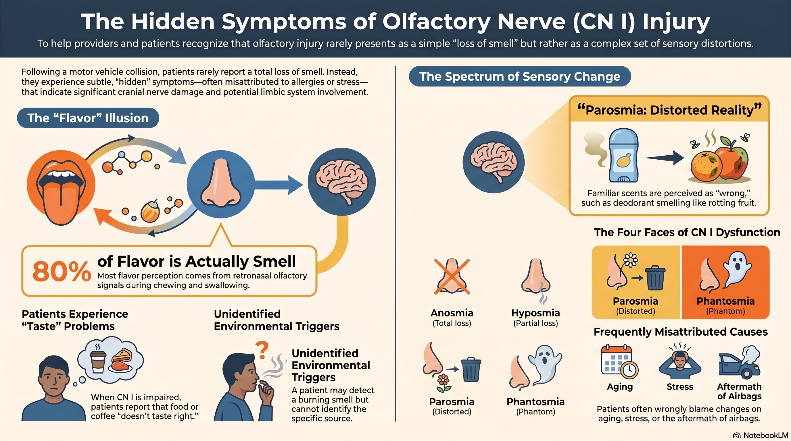
What patients more commonly report is a cluster of experiences that they do not connect to smell: food doesn’t taste the same anymore. Coffee doesn’t smell right. They can detect that something is burning but cannot identify what it is. Perfume that was familiar now smells different. They don’t enjoy meals the way they used to, and they can’t explain why.
Taste and smell are neurologically coupled. The experience of flavor is approximately 80 percent olfactory — what we perceive as the “taste” of food is largely the retronasal olfactory signal that occurs when aromatic compounds reach the olfactory epithelium during chewing and swallowing. When olfactory nerve function is impaired, the patient does not experience this as a smell problem. They experience it as a taste problem, or a food problem, or a generalized loss of pleasure in eating that they may not think to mention in a chiropractic or orthopedic visit.
There are also patients who develop parosmia — a distortion of smell in which familiar scents are perceived as unpleasant or wrong — rather than simple loss. A patient who reports that their partner’s deodorant now smells like rotting fruit, or that their own shampoo smells like chemicals, is not experiencing a minor sensory quirk. They are experiencing a measurable dysfunction in the olfactory pathway, with a specific and documentable presentation.
The full range of post-collision olfactory presentations includes anosmia (complete loss of smell), hyposmia (partial loss), parosmia (distorted smell), and phantosmia (smell of things that are not present). Each of these has clinical significance, each can be tested, and each has specific ICD-10 documentation pathways.
The patients who have these experiences rarely connect them to the collision without being asked. They have adapted. They have attributed the changes to aging, to stress, to a cold they had months ago. When a provider asks directly — “Since the accident, has your sense of smell changed? Do foods taste different?” — the frequency of positive responses is clinically significant and routinely surprising to providers who ask for the first time.
The symptom has been present since the crash. The question had never been asked.
The Examination Gap: What “Cranial Nerves Intact” Actually Means
A comprehensive cranial nerve examination covers twelve distinct systems. A complete exam generates up to 97 possible positive findings. It includes visual acuity and visual fields (CN II), extraocular movements (CN III, IV, VI), facial sensation (CN V), facial symmetry and motor function (CN VII), hearing and vestibular function (CN VIII), palatal movement and gag reflex (CN IX, X), shoulder and neck strength (CN XI), and tongue movement and symmetry (CN XII).
It also includes, as the first item in the sequence, olfaction — Cranial Nerve I.
The notation “cranial nerves 2 through 12 grossly intact” does not mean that all cranial nerves were examined and found intact. It means that cranial nerves 2 through 12 were assessed — and that CN I was either not tested or not documented. The phrase is so widely used that many providers write it as a template notation without performing the full examination it implies. This creates a documentation gap that serves no one: not the patient, not the provider, and certainly not the clinical record.
The omission has a specific effect in medicolegal contexts. When a defense expert reviews the clinical record and finds “cranial nerves 2-12 grossly intact” without a documented CN I assessment, there is no finding to dispute — but there is also no documented normal finding that would protect the provider’s examination from scrutiny. The correct documentation is explicit: either “CN I tested with [scent]; patient correctly identified/did not correctly identify the scent. CN I intact/impaired” or a specific explanation of why olfactory testing was deferred.
The reason CN I is omitted is not technical complexity. The test requires a cotton ball or familiar scented object — coffee, mint, citrus — held below the patient’s nostril with the other nostril occluded. The patient identifies the scent. The result is documented. This takes thirty seconds per side. It requires no specialized equipment, no referral, and no training beyond knowing that the test exists and that it should be performed.
The reason it is omitted is habit. The template was written without CN I. The attending clinician learned without emphasis on CN I. The training covered the visual, auditory, and motor cranial nerves with far more depth than the olfactory. The result is that the most commonly injured cranial nerve in motor vehicle collisions is systematically excluded from the examinations of the patients most likely to have injured it.
Closing that gap does not require new equipment. It does not require a specialist referral or additional training. It requires a question — “does this smell right to you?” — and the knowledge that the answer is clinically and legally significant.
The Clinical Protocol: Testing CN I and Documenting the Findings
The clinical protocol for olfactory nerve assessment in post-collision patients is straightforward and can be implemented immediately with materials already present in any clinical setting.
The first step is identifying appropriate test scents. The scents used must be recognizable, non-irritating, and sufficiently distinct. Common choices include coffee grounds, peppermint oil or a mint candy, citrus peel or citrus essential oil, vanilla extract, and cinnamon. A critical exclusion: do not use ammonia or other trigeminal irritants. These are detected via CN V — the trigeminal nerve — not CN I. A patient with complete olfactory loss can detect ammonia because it bypasses the olfactory pathway and activates trigeminal pain receptors. Using ammonia as a test scent would produce a false negative and is a significant clinical error.
The second step is testing each side independently. The patient occludes one nostril. The test scent is presented below the open nostril at a distance of approximately two centimeters, without allowing the scent container to contact the skin. The patient is asked to sniff normally and then identify the scent. The procedure is repeated on the other side. Bilateral testing matters because olfactory nerve damage may be asymmetric — unilateral injury is common, particularly in coup-contrecoup injuries where one hemisphere receives primary force, and would be missed entirely by testing both sides simultaneously.
The third step is documenting the findings with specificity. The clinical note should record the scent used, the side tested, and the patient’s response — both whether they detected any scent and whether they correctly identified it. A documentable notation reads: “CN I: patient tested bilaterally with peppermint oil. Right side: detected scent, unable to identify correctly. Left side: detected and correctly identified scent. Impression: right-sided hyposmia, possible olfactory nerve dysfunction, right CN I.” This notation is clinically meaningful, ICD-10 codeable, and legally documentable.
The fourth step is sequencing. Test CN I last in the cranial nerve examination. Standard clinical practice places olfactory testing at the end of the sequence to avoid olfactory fatigue — repeated scent exposure reduces sensitivity and can produce false results for hyposmia. Testing CN I last ensures the result reflects baseline olfactory function rather than examination-induced desensitization.
The fifth step is applying ICD-10 coding with specificity. Olfactory nerve dysfunction has dedicated ICD-10 codes for right and left sides. Anosmia and hyposmia each have distinct codes. Using the correct code — rather than a generic post-concussive symptom code — creates a specific documented injury with an impairment rating pathway. Under AMA Guides, olfactory loss qualifies for 1 to 5 percent whole-person impairment rating. That range is not large in isolation — but it is additive. A patient with cervical spine impairment, vestibular impairment, and olfactory impairment has three separately documented, separately ratable findings. Each adds to the total. None should be left untested.
Documentation language: “CN I tested bilaterally with [scent]. Right side: [patient detected / did not detect] scent; [correctly identified / unable to identify]. Left side: [patient detected / did not detect] scent; [correctly identified / unable to identify]. Impression: [intact bilaterally / right-sided hyposmia / anosmia] — possible olfactory nerve dysfunction. Referred for [neurological evaluation / formal olfactometry] as indicated.”
The Legal and Functional Weight of Olfactory Nerve Damage
Olfactory nerve injury carries weight in a personal injury case for reasons that are both anatomical and functional.
The anatomical weight is this: a documented CN I injury is objective evidence of brain injury. Not in the colloquial or exaggerated sense — in the specific, structural sense. The olfactory fila pass through the cribriform plate and synapse in the olfactory bulb. Damage to this system requires that enough mechanical force acted on the anterior cranial fossa during the collision to disrupt those fibers. This is not a soft tissue injury. It is a cranial nerve injury, and it is produced by the same category of force that produces concussion, intracranial hemorrhage, and diffuse axonal injury — though at a lower force threshold, which is precisely why it appears in collisions that produce no other radiographically visible brain injury.
The functional weight is this: olfactory loss changes daily life in ways that are well-documented and difficult to dispute. A patient who cannot smell cannot detect gas leaks, smoke, or spoiled food — a documented safety risk with direct daily consequences. A patient who experiences parosmia has had the sensory experience of eating permanently altered. A patient who has lost smell has lost the neurological infrastructure through which approximately 80 percent of flavor perception occurs. These losses are not trivial. They are documentable, they are measurable, and they persist.
Olfactory nerve damage, unlike many soft tissue injuries, does not reliably resolve with time. The olfactory fila have some regenerative capacity — olfactory receptor neurons are among the only neurons in the adult central nervous system capable of regeneration — but recovery is incomplete in a significant proportion of cases. Studies of post-traumatic olfactory loss consistently find that approximately one-third of affected patients never fully recover baseline olfactory function. This is a permanent injury with permanent functional impact, arising from a collision that may have produced no other finding on standard imaging.
In the medicolegal context, CN I injury has a specific effect on case value. It adds an objective finding to a case that may otherwise rest heavily on subjective complaint. It establishes that the collision was forceful enough to produce cranial nerve damage. It supports the patient’s reported cognitive and memory difficulties through anatomical proximity to the hippocampal-limbic system. And it opens the impairment rating pathway under AMA Guides in a way that is clean, specific, and difficult for a defense expert to challenge when the examination was properly performed and documented.
A defense expert who reviews a file containing a documented, bilaterally tested, correctly coded CN I injury with supporting ICD-10 designation and an AMA impairment rating is facing an objective finding. They can challenge the finding’s clinical significance, but they cannot make it disappear. They cannot substitute a retrospective normal for a documented positive.
The provider who tests and documents CN I has converted a patient complaint — “my memory hasn’t been right since the crash” — into a clinical finding with anatomical support, an ICD-10 code, and a permanent impairment pathway. That conversion is what complete post-collision care looks like.
“The provider who tests CN I has converted a patient complaint into a clinical finding. That conversion is what complete post-collision care looks like.”
Conclusion: The Test That Takes Thirty Seconds
Post-collision clinical evaluation is a process of finding everything that the collision did to a patient — not just what is visible on imaging, not just what fits into the musculoskeletal model, not just what the patient thought to mention when they walked in reporting a stiff neck.
The olfactory nerve sits at the intersection of the most common mechanism of cranial nerve injury in motor vehicle collisions and the most common cognitive complaint in post-collision patients. It is adjacent, by anatomy, to the memory structures that patients are struggling to identify as impaired. It is excluded, by habit, from the examinations of the patients most likely to have injured it. And it can be tested, by any clinician, in thirty seconds, with a familiar scent and a documented response.
The most commonly injured cranial nerve in motor vehicle collisions is also the one most commonly untested. That is not an immutable clinical reality. It is a correctable examination gap — one that, once closed, produces objective findings, supports patient-reported symptoms with anatomical specificity, creates a documented impairment pathway, and ensures that the clinical record reflects what actually happened to the patient.
When a patient presents with memory problems after a crash, the question is not whether to investigate — it is which system to examine first. The olfactory nerve answers that question in thirty seconds. Its proximity to the hippocampus and entorhinal cortex makes a positive finding more than just a smell disorder. It is a gateway finding that confirms brain exposure to sufficient force, supports the cognitive complaints the patient has been describing, and opens the complete documentation pathway the case requires.
The test takes thirty seconds. The finding lasts the lifetime of the case.
References
- Costanzo RM, Becker DP. Smell and taste disorders in head injury and neurosurgery patients. In: Meiselman HL, Rivlin RS, eds. Clinical Measurement of Taste and Smell. New York: Macmillan; 1986.
- Sumner D. Post-traumatic anosmia. Brain. 1964;87(1):107–120.
- Fortin A, Lefebvre MB, Ptito M. Traumatic brain injury and olfactory deficits: the tale of two smell tests. Brain Injury. 2010;24(1):27–33.
- Zusho H. Posttraumatic anosmia. Archives of Otolaryngology. 1982;108(2):90–92.
- American Medical Association. Guides to the Evaluation of Permanent Impairment, 6th ed. Chicago: AMA; 2008.