When Normal Isn’t the Whole Story: The Hidden Injuries That Imaging Misses After a Car Accident
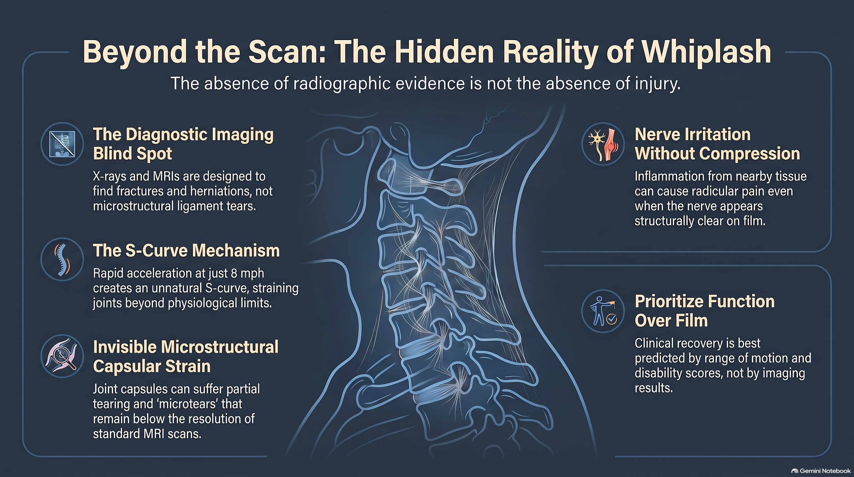
Between 15 and 40 percent of people involved in rear-end collisions will develop chronic symptoms that persist longer than one year. In the majority of these cases, the imaging is normal. The bones are aligned. The discs are intact. The radiologist’s report uses phrases like “no acute findings,” “age-appropriate changes,” and “within normal limits.” The patient leaves with that report in hand, and for many of them, it becomes a source of confusion that compounds the injury itself. If nothing is wrong, why does everything hurt? If the scan is clear, what is there to treat?
The answer requires understanding something the imaging does not show: that the tissues most commonly injured in low-speed collisions are not the tissues that imaging is designed to detect. The gap between the patient’s experience and the radiographic record is not an illusion or an exaggeration. It is a consequence of using the wrong diagnostic tool for the job — and then treating its silence as evidence.
This article examines what imaging can and cannot see after a motor vehicle collision, what the research says about the injuries it routinely misses, and why the clinical standard for evaluating post-collision patients must extend well beyond what appears on film.
What Imaging Was Designed to Find — and What It Wasn’t
X-ray was developed to visualize dense structures — principally bone. It is highly effective at identifying fractures, dislocations, and gross malalignment of the vertebral column. In the context of a motor vehicle collision, cervical X-ray rules out the serious but relatively uncommon injuries: odontoid fractures, bilateral facet dislocations, burst fractures of the vertebral body. These are the injuries emergency physicians are appropriately screening for when they order imaging after a crash.
MRI expanded the diagnostic window considerably. It can visualize soft tissue — intervertebral discs, the spinal cord, and with high-field equipment, portions of the musculature. A herniated disc compressing a nerve root will appear on MRI. Spinal cord contusion will appear on MRI. Large extradural hematomas will appear on MRI.
What neither modality was designed to detect — and what both routinely miss — is the category of injury most common in low-speed rear-end collisions: partial tearing of spinal ligaments, capsular disruption at the facet joints, diffuse muscle fiber damage without complete rupture, and nerve root or dorsal root ganglion irritation without structural compression.
These injuries are real. They are measurable by other means. They are associated with significant and sometimes permanent functional limitation. But they exist below or outside the threshold of visibility on standard imaging, which means that a “normal scan” after a rear-end collision does not mean no injury occurred. It means the imaging did not detect the kind of injury that standard imaging is equipped to detect.
This distinction matters enormously for patients, clinicians, and anyone evaluating the legitimacy of a collision claim. The absence of radiographic evidence is not the same as the absence of injury. It is the absence of radiographic evidence.
The Biomechanics of Low-Speed Impact
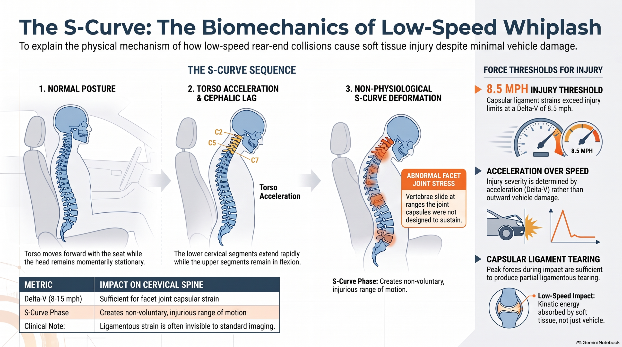
To understand why low-speed collisions produce significant soft tissue injury without bony damage, it is necessary to understand what happens to the cervical spine during a rear-end collision at speeds that might seem inconsequential.
Research using instrumented volunteers, crash test dummies, and cadaveric specimens has established that the forces acting on the cervical spine during a rear-end collision do not scale linearly with vehicle speed. The critical variable is not speed but acceleration — specifically, the rate of change of velocity, often expressed as delta-V, and the duration over which that acceleration is applied to the occupant’s body.
In a low-speed rear-end collision — commonly defined as a delta-V between 8 and 15 miles per hour — the vehicle structure absorbs much of the kinetic energy through crumple zones and bumper deformation. The occupant’s body, however, is carried by the seatback and transmitted upward toward the head. This transmission is not uniform. The torso moves first, forward with the vehicle. The head, connected to the torso by the cervical spine, follows — but with a lag. This lag creates a brief, transient S-curve deformation of the cervical spine: the lower segments extend rapidly while the upper segments are still in relative flexion.
This S-curve configuration — documented extensively by Panjabi and colleagues using cervical spine specimens and high-speed cinematography — does not occur in any normal voluntary movement. It creates a pattern of facet joint stress that the cervical spine is not designed to accommodate. The inferior articular facets of the upper vertebrae slide posteriorly relative to the superior facets of the lower vertebrae at velocities and through ranges that the joint capsule was not designed to sustain.
The peak forces at the facet joint capsule during a low-speed rear-end collision have been measured, in volunteer and cadaver studies, at levels sufficient to produce capsular strain and partial tearing — even in the absence of bony injury and even when vehicle damage is minimal. A 1998 study by Pearson and colleagues demonstrated that facet joint capsule strains during simulated rear-end collisions exceeded the injury threshold for capsular ligament tissue at delta-V values as low as 8.5 miles per hour.
The vehicle may look fine. The X-ray may look fine. The facet joint capsule may not be fine.
“The absence of radiographic evidence is not the same as the absence of injury. It is the absence of radiographic evidence.”
Ligamentous Injury: The Invisible Architecture
The cervical spine is stabilized by a complex network of ligaments whose primary function is to constrain movement within the normal physiological range and protect the spinal cord and nerve roots from excessive displacement. These ligaments — the anterior longitudinal ligament, the posterior longitudinal ligament, the ligamentum flavum, the interspinous ligaments, the capsular ligaments of the facet joints, and the specialized upper cervical ligaments including the alar and transverse ligaments — are composed primarily of collagen fibers organized in highly structured arrays.
When stretched beyond their physiological limit, these ligaments do not behave like rubber bands. They do not simply elongate and return. Collagen fibers have a characteristic load-deformation curve: they tolerate elastic deformation up to a point, after which individual fibers begin to rupture sequentially. Partial tearing of a ligament — sometimes called a sprain or microtear — does not produce the complete disruption visible on imaging. It produces a zone of disrupted collagen architecture within an otherwise intact-appearing structure. Standard MRI cannot reliably detect this.
Marios Panjabi’s biomechanical research introduced the concept of the “neutral zone” — the range of spinal motion near the neutral position within which there is minimal internal resistance from the passive stabilizing structures. In a healthy spine, the neutral zone is narrow because the ligamentous and capsular structures provide effective restraint throughout the range of motion. When those structures are partially torn, the neutral zone widens. The spine moves farther than it should before the passive restraints engage. This widening is the biomechanical signature of ligamentous instability — and it is not visible on static imaging. It can be detected on stress radiographs and with dynamic functional testing, but these are rarely performed in standard emergency or urgent care evaluations.
The alar ligaments — two stout cords connecting the tip of the dens to the inner surface of the occipital condyles — stabilize the atlanto-axial joint against excessive rotation and lateral bending. Alar ligament injury has been identified using high-field MRI (3 Tesla), but standard 1.5 Tesla MRI frequently fails to visualize partial tearing. A 2008 study by Kaale and colleagues compared alar ligament findings on 3T MRI in patients with chronic whiplash-associated disorders to asymptomatic controls. They found significantly higher rates of alar ligament signal change in the whiplash group — signal changes consistent with internal disruption. These changes were not visible on standard clinical MRI, and they were not produced by placebo. They were produced by injury.
The capsular ligaments of the cervical facet joints present a similar problem. They are visible on MRI in their intact form, but partial capsular tears — the type of injury produced by the strain patterns documented in volunteer collision studies — are below the resolution of clinical MRI sequences. The joint looks normal. The capsule is not.
This is not a theoretical limitation. It is a documented, replicated discordance between imaging findings and histopathological evidence of tissue damage. Post-mortem studies of individuals who died from causes unrelated to their motor vehicle collision — but who had documented chronic whiplash symptoms prior to death — have found macroscopic and microscopic evidence of ligamentous disruption in the absence of any corresponding abnormality on ante-mortem imaging. The injury was there. The imaging did not see it.
Microtears, Muscle Pathology, and the Limits of Conventional MRI
The cervical musculature is vulnerable to a distinct category of injury in rear-end collisions — one that has been documented using advanced imaging modalities but is invisible on conventional clinical MRI.
Rapid eccentric loading — the type of force applied to the cervical extensors as the head whips forward relative to the thorax during the initial phase of a rear-end collision — is a well-established mechanism for intramuscular injury. Eccentric loading produces microtears within muscle fiber bundles, small foci of intramuscular hemorrhage, and disruption of the connective tissue scaffolding that organizes muscle architecture. These changes are not macroscopic tears. They are distributed, diffuse injuries to the microstructure of the muscle belly.
On standard 1.5T MRI, these changes do not produce reliable signal abnormality. The resolution is insufficient to detect diffuse intramuscular pathology of this type unless it is associated with frank hemorrhage or complete rupture — neither of which typically occurs in low-speed collisions.
However, advanced imaging modalities — including T2 mapping, diffusion tensor imaging (DTI), and high-resolution ultrasound — have demonstrated consistent abnormalities in the cervical musculature of patients with whiplash-associated disorders, even when standard MRI appears normal.
Research by Elliott and colleagues, using fat-fraction analysis on MRI, documented increased fatty infiltration of the cervical multifidus in patients with chronic whiplash-associated disorders compared to healthy controls. Fatty infiltration is a late consequence of muscle injury — it represents the replacement of functional contractile tissue with non-contractile fat, a process that reflects underlying structural damage that was never visible on the original clinical imaging. The finding was not present in the acute phase. It developed over months of abnormal muscle use following injury — the chronic consequence of a microstructural injury that never appeared on the scan.
This finding carries significant clinical implications. The multifidus is the primary deep stabilizer of the cervical spine. Its atrophy and fatty infiltration reflect not only the direct injury to the muscle tissue but also the secondary consequence of proprioceptive disruption: when the deep cervical stabilizers reduce their activity in response to sensory disruption, they undergo disuse-related changes that can be measured years after the initial collision — changes directly traceable to an injury event that produced no detectable finding on the scan taken in the emergency department.
Nerve Irritation Without Structural Compression
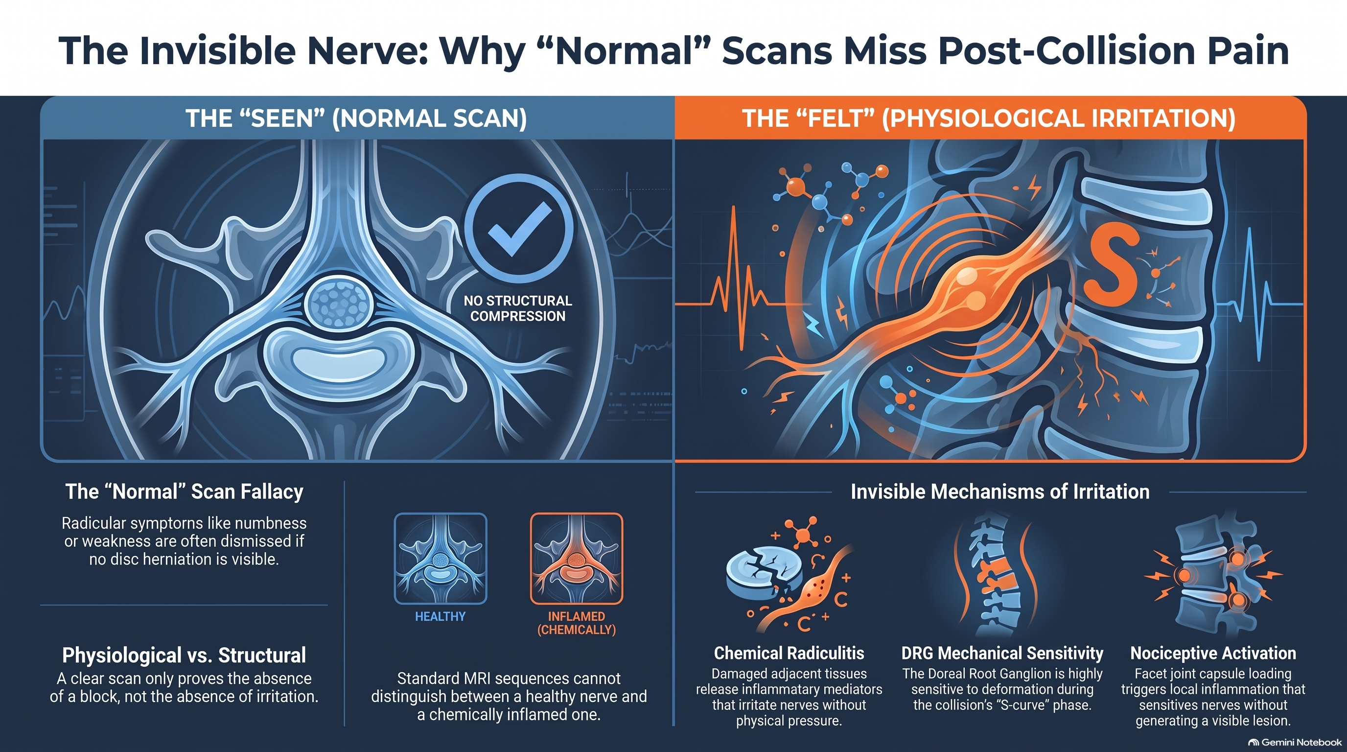
Radicular symptoms — pain, numbness, or weakness in an arm or hand following a cervical injury — are among the most commonly dismissed findings in post-collision patients with normal imaging. The logic is intuitive: if there is no disc herniation pressing on a nerve root, there is no reason for radicular symptoms. The scan is normal, therefore the nerve is fine.
This logic misunderstands the mechanism by which nerve roots and dorsal root ganglia generate symptoms.
The dorsal root ganglion (DRG) — the cluster of sensory neuron cell bodies located in the intervertebral foramen — is one of the most mechanically sensitive structures in the nervous system. Unlike nerve tissue within the dural tube, the DRG has a relatively poor blood-nerve barrier, making it highly susceptible to chemical irritation from inflammatory mediators released by damaged adjacent tissues. It is also directly vulnerable to mechanical deformation from forces transmitted through the foramen during the collision’s S-curve phase.
Chemical radiculitis — radicular symptoms produced not by structural compression but by chemical irritation of the DRG from inflammatory mediators released by adjacent damaged tissue — produces symptoms clinically indistinguishable from disc herniation radiculopathy. Pain radiates in a dermatomal pattern. Deep tendon reflexes may be reduced. Electrodiagnostic testing (EMG and nerve conduction studies) may be normal because the nerve’s electrical conduction is intact — only its sensitivity to chemical stimulation has been altered.
A normal MRI cannot distinguish between a nerve root that is completely unaffected and a dorsal root ganglion that is inflamed by mediators released from an adjacent capsular tear. Both look identical on standard sequences. The difference is physiological, not structural. It is detectable by the patient’s symptoms, by the clinician’s examination findings, and by the pattern of response to appropriate treatment — but not by the imaging.
Research by Cavanaugh and colleagues has established that facet joint capsule loading — at levels consistent with low-speed rear-end collisions — activates the nociceptive afferents within and around the capsule, including fibers that project to adjacent DRGs. This activation produces a cascade of local inflammation that can sensitize the DRG without producing any compressive lesion and without generating any MRI-visible change. The nerve is irritated. The scan is clear.
Chemical radiculitis produces arm pain, numbness, and reflex changes that are clinically indistinguishable from disc compression — but it does not appear on MRI and EMG is typically normal. The nerve is irritated by inflammation from adjacent capsular injury, not compressed by a structural lesion.
The Clinical Standard: Function Over Film
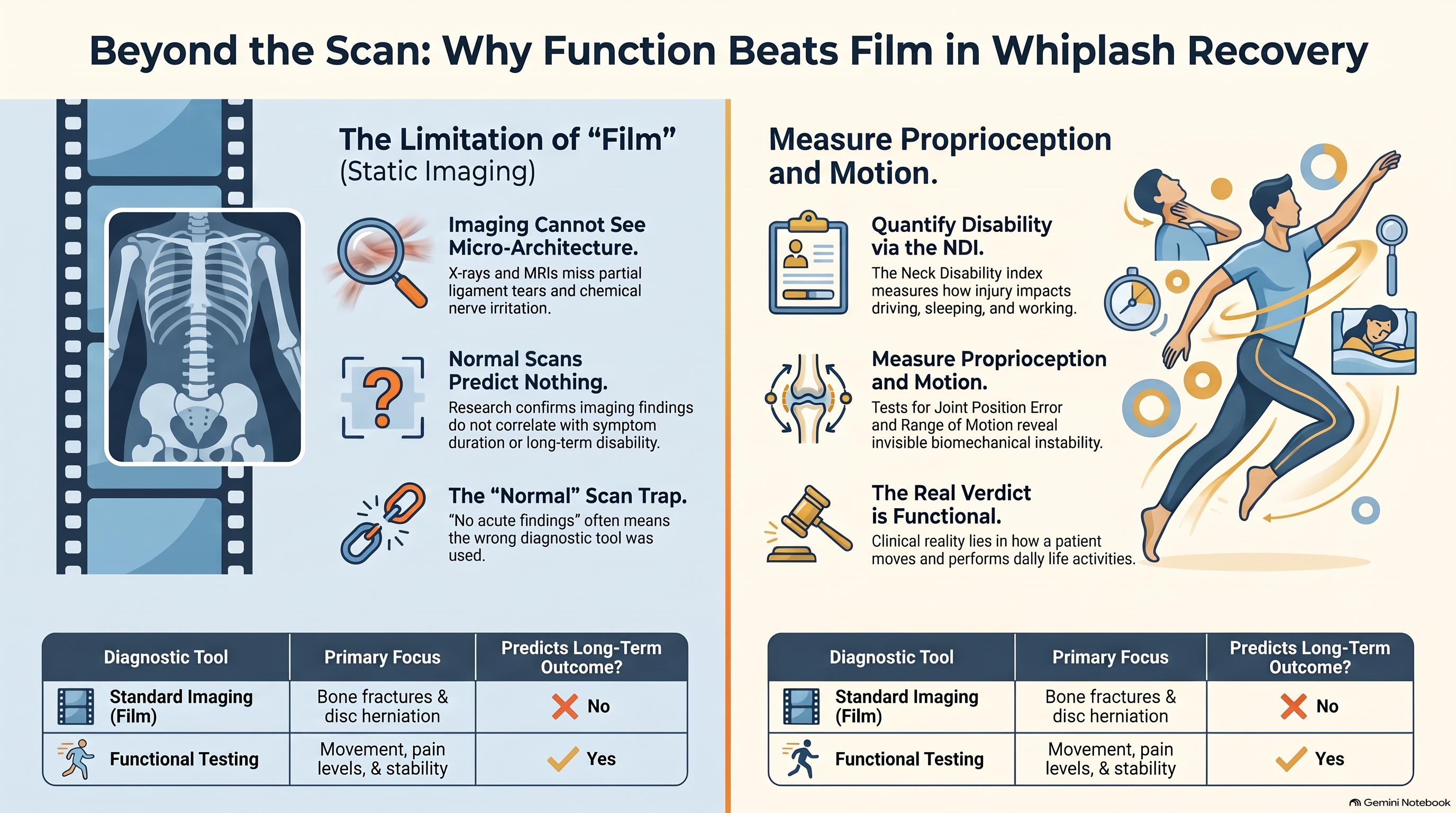
Given the limitations of standard imaging in detecting the injuries most common in post-collision patients, the appropriate clinical standard for evaluating these patients is not radiographic — it is functional.
The instruments developed and validated for this purpose measure what the imaging cannot: how the patient moves, how accurately the neck’s sensory system reports position, how much pain interferes with daily function, and how the patient’s capacity to perform basic activities has changed since the injury.
The Neck Disability Index (NDI), a ten-item patient-reported outcome measure, captures interference with activities including personal care, lifting, reading, driving, sleeping, and work. Its reliability and validity in post-whiplash populations have been extensively documented. Patients with normal imaging frequently score in the moderate to severe disability range on the NDI — and those scores predict long-term outcomes more accurately than any imaging finding.
The joint position error test — in which the patient closes their eyes, moves the head away from neutral, and attempts to return to the starting position — provides a direct measure of proprioceptive accuracy. Patients with whiplash-associated disorders show significantly elevated joint position error compared to healthy controls, reflecting the disruption of cervical muscle spindle function. This disruption is not visible on any clinical imaging modality. It is measurable only through functional testing.
Cervical range of motion — measured with an inclinometer or specialized goniometer — quantifies the functional consequence of the injury. Restriction of range of motion in post-collision patients correlates with symptom severity, disability scores, and long-term recovery trajectory. It is directly measurable in the clinical setting. It does not require imaging.
The Visual Analogue Scale or Numeric Pain Rating Scale captures pain intensity — the cardinal symptom of injury, regardless of radiographic appearance. Pain is a physiological event that occurs in the nervous system, not in the X-ray. Its presence is clinically valid independent of imaging findings.
The clinician who evaluates a post-collision patient using only imaging — who reads “no acute findings” and concludes that no acute injury occurred — has used the wrong tool for the clinical question. The right tool is the one that measures what was actually injured: function.
The Medico-Legal Consequence of Radiographic Silence
The clinical reality described in this article collides directly with a persistent pattern in personal injury claims management: the use of a “normal scan” to impute that no injury occurred, that no treatment is necessary, and that no compensation is warranted.
This pattern is medically indefensible — but it is pervasive. Insurance adjusters, independent medical examiners employed by defense firms, and opposing counsel routinely cite negative imaging as evidence that a claimant’s symptoms are exaggerated, manufactured, or attributable to pre-existing conditions. The logic appears straightforward: if nothing shows on the scan, nothing was injured.
The research literature does not support this logic. It directly contradicts it.
A 2003 systematic review by Scholten-Peeters and colleagues examined the predictors of chronic disability following whiplash injury across 29 studies. Radiographic findings were not among the significant predictors of long-term outcome. The factors that predicted chronicity were neurological symptoms at presentation, high initial pain intensity, and psychological factors — not the presence or absence of imaging abnormality. The scan did not predict who recovered and who did not. It predicted nothing clinically useful about long-term outcome.
A 2002 review by Hartling and colleagues, examining the evidence base for whiplash prognosis, reached a similar conclusion: imaging findings were not associated with symptom duration or functional outcome in the majority of included studies. The patients who developed chronic disability were not, as a group, the patients with abnormal imaging. They were the patients with high initial pain scores, limited cervical range of motion, and elevated psychological distress — all of which are functional measures, not radiographic ones.
Clinical documentation standard: Record the NDI score, cervical range of motion, joint position error, and pain ratings for every post-collision patient — regardless of imaging findings. These functional measures are the evidentiary record that reflects the actual clinical reality of the injury.
The clinician’s responsibility in this context is clear: document function comprehensively. Record the NDI score. Record the range of motion measurements. Record the joint position error results. Record the pain scores. Record the specific activities the patient can no longer perform — and the specific activities they can. This documentation creates the evidentiary record that reflects what the imaging cannot: the actual clinical reality of the patient’s injury.
When a patient with a normal scan cannot drive more than fifteen minutes without neck pain, cannot sleep through the night without waking in spasm, and cannot return to their physical work — that is a clinical finding. It is measurable, documented, and reproducible. It does not require radiographic correlation. It requires a clinician willing to look beyond the image.
Conclusion
The phrase “your scan is normal” is one of the most consequential things a clinician can say to a post-collision patient — and one of the most frequently misunderstood. It is not a verdict. It is a statement about what one diagnostic tool, used for a specific purpose, was able to detect in a specific tissue at a specific moment in time.
The injuries that follow low-speed rear-end collisions — ligamentous microtears, capsular disruption, intramuscular pathology, proprioceptive deficit, and chemical nerve irritation — are not reliably detectable by standard imaging because they are not the injuries that standard imaging was designed to find. Their existence is not a matter of debate. The biomechanical research that documents them, the histopathological evidence that confirms them, and the functional outcome literature that traces their clinical consequences are among the most robustly replicated bodies of evidence in musculoskeletal medicine.
The real verdict after a motor vehicle collision does not lie in film. It lies in function: in how the patient moves, what the sensory system reports, how daily life has been altered, and whether the tissues responsible for stability and position sense have been restored to their pre-injury state. These are measurable. These are treatable. And these are what the clinical encounter must be organized around — not the reassuring silence of a scan that was never designed to speak to the question being asked.
References
- Panjabi MM, Cholewicki J, Nibu K, et al. Simulation of whiplash trauma using whole cervical spine specimens. Spine. 1998;23(1):17–24.
- Pearson AM, Ivancic PC, Ito S, Panjabi MM. Facet joint kinematics and injury mechanisms during simulated whiplash. Spine. 2004;29(4):390–397.
- Kaale BR, Krakenes J, Albrektsen G, Wester K. Head position and impact direction in whiplash injuries: associations with MRI-verified lesions of ligaments and membranes in the upper cervical spine. Journal of Neurotrauma. 2005;22(11):1294–1302.
- Elliott JM, Pedler AR, Kenardy J, et al. The temporal development of fatty infiltrates in the neck muscles following whiplash injury. PLoS ONE. 2011;6(8):e21194.
- Cavanaugh JM, Lu Y, Chen C, Kallakuri S. Pain generation in lumbar and cervical facet joints. Journal of Bone and Joint Surgery. 2006;88(Suppl 2):63–67.
- Scholten-Peeters GG, Verhagen AP, Bekkering GE, et al. Prognostic factors of whiplash-associated disorders: a systematic review of prospective cohort studies. Pain. 2003;104(1–2):303–322.
- Hartling L, Brison RJ, Ardern C, Pickett W. Prognostic value of the Quebec Classification of Whiplash-Associated Disorders. Spine. 2001;26(1):36–41.