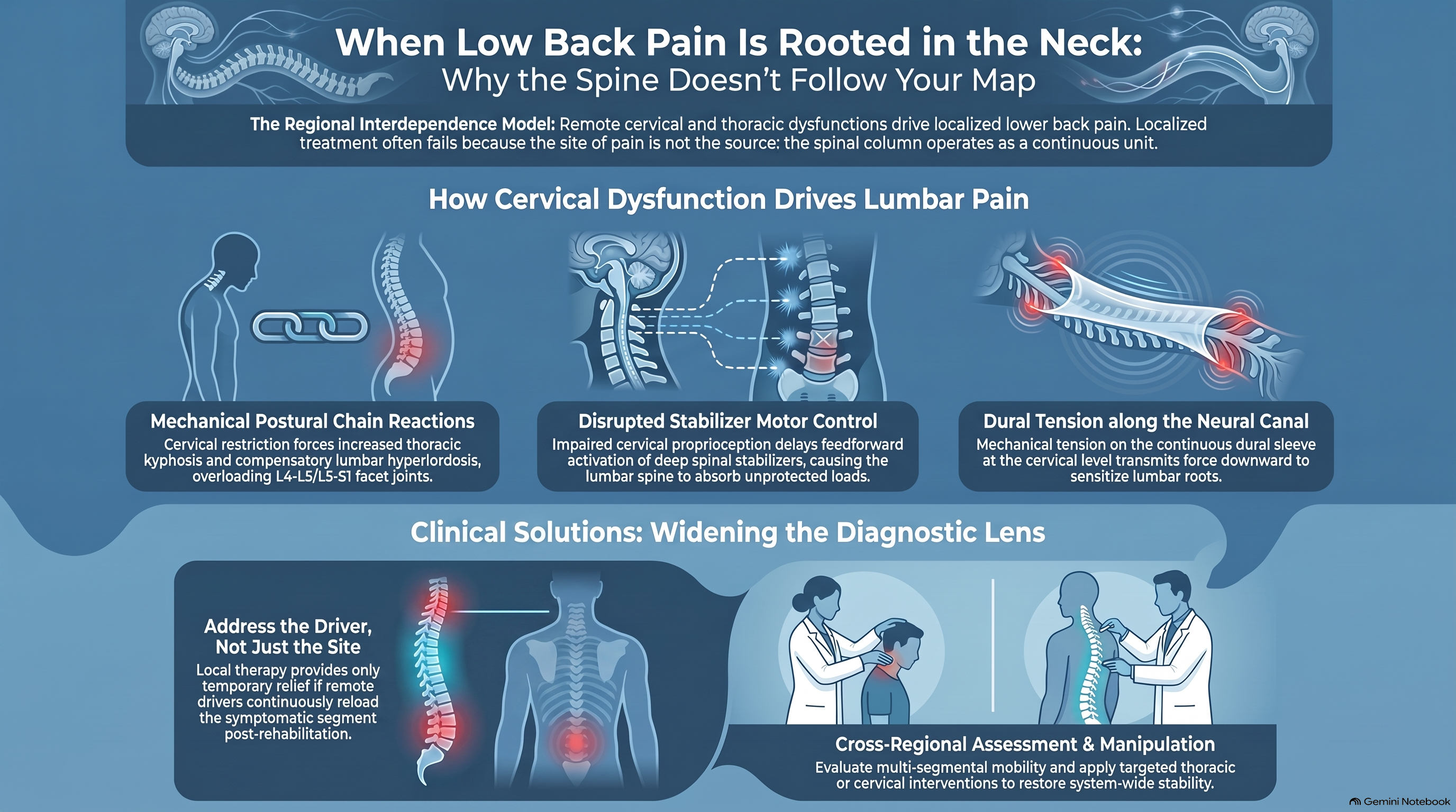
When Low Back Pain Is Rooted in the Neck: Why the Spine Doesn’t Follow Your Map
Your low back has been treated for six weeks. The manual therapy is correct. The exercises are appropriate. The diagnosis is documented. The imaging confirms degeneration at L4-5. And nothing is working.
The problem may not be in your low back.
One of the most consistently underrecognized patterns in spinal pain management is regional interdependence — the clinical phenomenon in which pain at one region of the spine is driven, maintained, or modulated by dysfunction at a remote region. The lumbar spine can be symptomatic because of a cervical or thoracic abnormality. The cervical spine can be symptomatic because of a lumbar or pelvic problem. A clenched jaw can propagate tension downward through the thoracic extensors to the pelvic floor. A rigid thoracic spine can force compensatory hypermobility at both cervical and lumbar segments, producing symptoms at those endpoints while the true restriction remains untouched in the middle.
Pain’s geography is deceptive. The site of pain is not always the source of pain. And the clinician who treats only where it hurts — without mapping the interconnected architecture of the spinal column as a single functional unit — will manage symptoms without resolving the underlying driver.
This article examines the mechanisms by which cervical dysfunction produces lumbar symptoms, the reverse relationship by which lumbar pathology drives cervical pain, the neurological and biomechanical pathways that connect them, and what this means for clinical assessment and treatment planning.
The Regional Interdependence Model: A Spine That Doesn’t Know Its Segments
The concept of regional interdependence — formalized in physical therapy and manual medicine literature by Wainner, Whitman, and colleagues — proposes that musculoskeletal impairments in one region of the body can directly contribute to symptoms and impairments in another, apparently unrelated region. This is not a theoretical abstraction. It is a clinically documented, biomechanically grounded phenomenon with direct implications for assessment and treatment outcomes.
The spine is not organized into the discrete segments that clinical specialization implies. Radiologists image by region. Surgeons operate by region. Insurance forms are coded by region. But the biomechanical and neurological systems that govern spinal function do not observe these divisions. The deep stabilizing musculature — the multifidus, the rotatores, the semispinalis — functions as a continuous system from the occiput to the sacrum. The thoracolumbar fascia connects the lumbar extensors to the cervical system through the posterior chain. The dura mater, the connective tissue sleeve enveloping the spinal cord, is mechanically continuous throughout the canal. Neural tension at the cervical level transmits force along the neural canal and can be perceived as symptoms at remote lumbar or lower extremity levels.
When one region of this continuous system is dysfunctional — through injury, chronic guarding, deconditioning, or joint restriction — the compensatory demands placed on adjacent and remote regions increase. The system maintains function through compensation. But compensation has a cost: the regions absorbing the compensatory load become overloaded, symptomatic, and eventually structurally compromised. The patient presents with lumbar pain. The source of the compensatory demand is in the thoracic spine — or the cervical spine — or the sacroiliac joint. Treatment at the lumbar level reduces symptoms temporarily. The compensatory demand reloads the lumbar segment as soon as rehabilitation ends. And the patient cycles.
How the Cervical Spine Drives Lumbar Symptoms
The mechanisms by which cervical dysfunction produces or contributes to lumbar symptoms are multiple and interconnected.
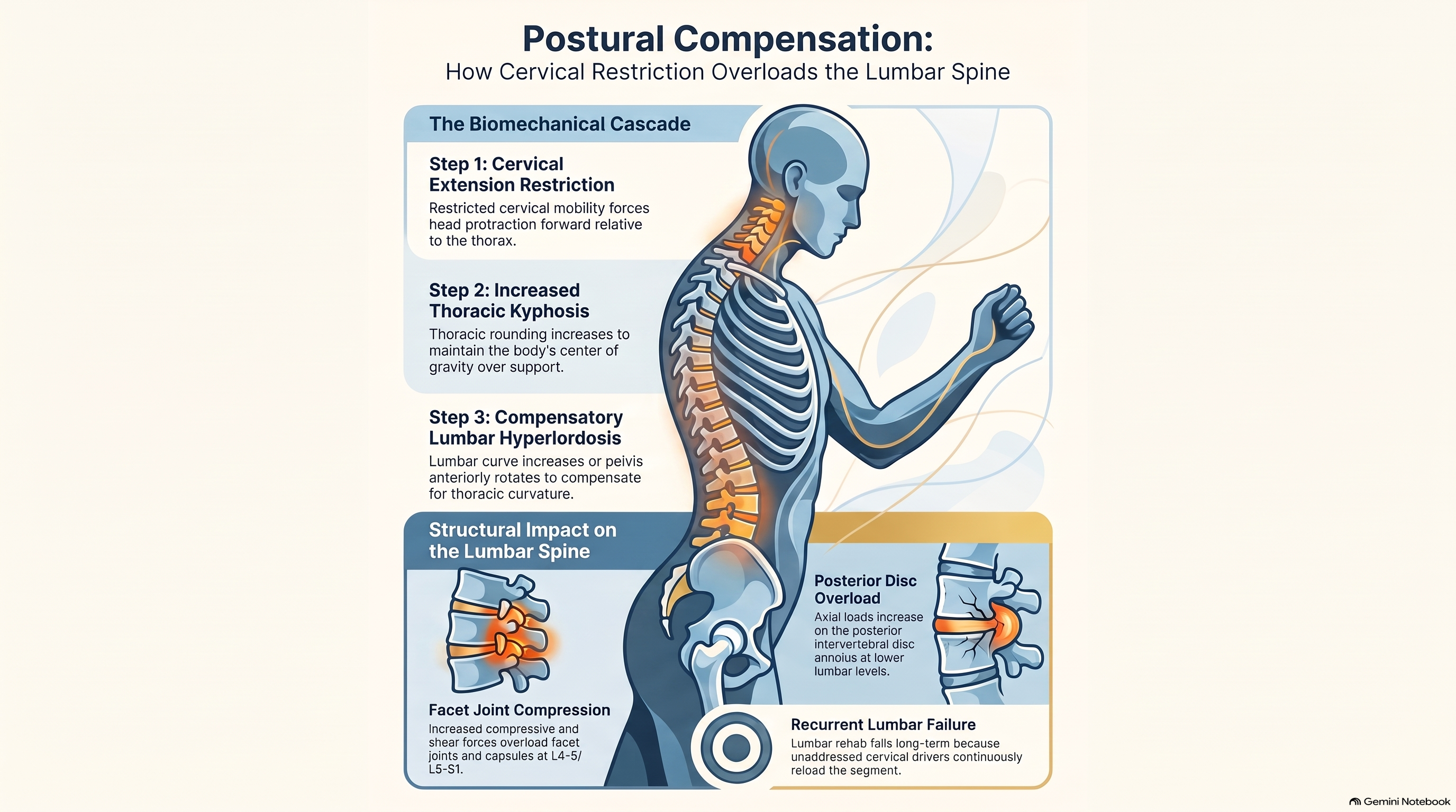
Postural compensation through the thoracic spine. The most direct pathway is mechanical. A cervical spine with restricted extension — whether from post-collision capsular tightness, chronic segmental restriction, or protective muscle guarding — alters head position relative to the thorax. The head moves forward into the protracted position. To maintain the center of gravity over the base of support, the thoracic kyphosis increases. To compensate for increased thoracic kyphosis, the lumbar lordosis must increase correspondingly, or the pelvis anteriorly rotates, or both. This compensatory lumbar hyperlordosis increases compressive and shear forces at the posterior elements — facet joints and capsules at L4-5 and L5-S1 — and increases the axial load on the posterior intervertebral disc annulus at those levels. The cervical restriction drives the thoracic posture. The thoracic posture drives the lumbar loading. The lumbar segment fails under the compensatory load.
Proprioceptive disruption and motor control changes. The cervical spine, particularly the suboccipital region, provides the highest density of proprioceptive input per unit area of any region of the spinal column. When cervical proprioception is disrupted — through joint capsule injury, deep cervical muscle dysfunction, or vestibular co-impairment — the central nervous system’s ability to coordinate axial motor control is compromised throughout the spine. Research by Hodges and Richardson documented that in healthy subjects, the deep spinal stabilizers — the multifidus and transversus abdominis — activate in anticipation of limb movement, preparing the spine before the peripheral load arrives. This feedforward activation is coordinated by central motor programs that depend, in part, on accurate cervical proprioceptive input. When that input is degraded, the feedforward activation is delayed or absent. The lumbar spine absorbs unprotected loads. Over time, the cumulative effect of repeated unprotected loading produces lumbar segment pathology — even in the absence of any direct lumbar injury.
Neural tension and dural continuity. The dural sleeve is mechanically continuous from the cranium to the sacrum. Increased neural tension at the cervical level — through foraminal narrowing, capsular adhesion, or dural tethering following cervical disc pathology — transmits mechanical load along the neural canal. This can produce referral of symptoms into the lower extremities through sensitized lumbar nerve roots, even when those roots themselves show no structural compromise. The mechanism is central sensitization combined with dural mechanical transmission: the sensitized lumbar nerve root, receiving heightened nociceptive input from the central sensitization established by the cervical pathology, produces symptoms that map to lumbar or lower extremity distributions.
How Lumbar Pathology Produces Cervical Pain
The reverse relationship is equally well-documented and equally underrecognized.
Antalgic posture and cervical loading. A patient with significant lumbar pain adopts a characteristic posture: reduced lumbar lordosis, forward trunk lean, and increased thoracic kyphosis. This posture, maintained over time, places the cervical spine in a chronically protracted position — the head forward, the cervical extensors in sustained contraction, the cervical facet joints compressed posteriorly. The sustained cervical extensor contraction produces myofascial pain at the suboccipital region, the posterior cervical musculature, and the upper trapezius. The patient reports neck pain and headache. The source is the lumbar antalgic posture. Treatment focused on the cervical spine will reduce the local symptoms without addressing the postural driver.
Sacroiliac dysfunction and lumbar-thoracic compensation. Sacroiliac joint dysfunction — whether from direct trauma, post-partum ligamentous laxity, or cumulative micro-trauma — produces a characteristic pattern of trunk compensation: reduced ipsilateral gluteus medius activation, increased contralateral quadratus lumborum activation, a lateral pelvic tilt, and compensatory scoliotic deviation through the thoracolumbar junction. This deviation propagates upward through the thoracic spine and is absorbed, in part, by increased segmental stress at the cervico-thoracic junction. Patients with chronic sacroiliac dysfunction frequently present with cervico-thoracic pain and restricted cervical rotation to one side — findings that persist until the sacroiliac driver is identified and addressed.
Central sensitization from lumbar sources. Chronic lumbar discogenic pain, facet joint pain, or sacroiliac pain produces central sensitization — an upregulation of the dorsal horn pain processing circuitry that lowers the threshold for nociceptive transmission throughout the sensitized spinal segments. When the lumbar sensitization is significant and sustained, the sensitized state can extend to adjacent and remote spinal levels. Cervical inputs that would be sub-threshold in a normal nervous system become symptomatic when the system is already sensitized from below. The patient reports both cervical and lumbar pain simultaneously, and treatment of either region alone produces only partial relief.
The Jaw, the Pelvis, and the Chain Reaction
The research literature on temporomandibular joint dysfunction provides one of the most striking examples of remote spinal influence: a clenched jaw can produce pelvic floor tension.
The connection is not metaphorical. The hyoid bone — suspended between the mandible above and the sternum and clavicle below — is a central node in the anterior cervical chain. The suprahyoid and infrahyoid muscles connect it upward to the floor of the mouth and the temporomandibular apparatus, and downward to the sternum, clavicle, and scapula. Chronic TMJ dysfunction or bruxism produces sustained activation of the suprahyoid musculature, which creates anterior tension on the hyoid. This anterior tension activates the infrahyoid muscles, producing tension at the sternum and anterior cervical fascia. The anterior cervical fascia is continuous with the thoracic fascia through the pericardium and mediastinal connective tissue. Fascial tension transmitted downward through this anterior chain reaches the thoracolumbar junction and the anterior abdominal wall, producing increased intra-abdominal pressure and reflexive pelvic floor activation.
The patient with chronic TMJ dysfunction, persistent cervical tension, and unexplained pelvic floor tightness is not presenting with three separate diagnoses. They are presenting with one continuous fascial chain under sustained anterior tension, expressed at three geographically separated points.
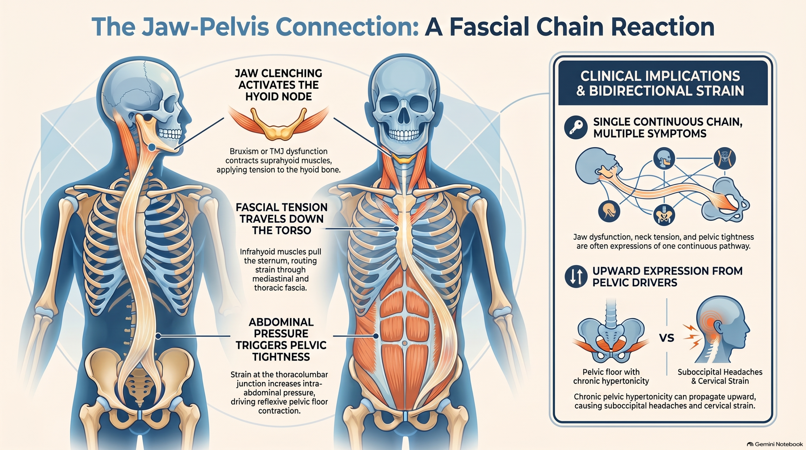
The same logic applies in reverse. A patient with chronic pelvic floor hypertonicity — from post-partum trauma, chronic stress activation, or sacroiliac dysfunction — may present with cervical tension and suboccipital headache as the upward expression of the same anterior chain dysfunction.
Clinical Implications: Widening the Diagnostic Lens
The clinical implication of regional interdependence is straightforward to state and genuinely difficult to implement: examination must not stop at the region of pain.
Every post-collision patient with cervical symptoms should have their thoracic mobility and lumbar posture assessed. Every patient with persistent lumbar pain unresponsive to local treatment should have their cervical range of motion, deep flexor activation, and thoracic extension measured. Every patient with TMJ symptoms should be assessed for cervical proprioceptive deficit and hyoid muscle tension. The regions are connected. The assessment must be too.
Specific clinical tests that cross regional boundaries include the slump test and straight leg raise, which assess neural tension through both lumbar and cervical contributions to the dural sleeve — cervical flexion sensitization in the slump test is a direct measure of dural continuity between cervical and lumbar levels. The cervical rotation lateral flexion (CRFL) test assesses first rib mobility and its contribution to cervico-thoracic junction restriction that can drive both cervical and upper extremity symptoms. The active straight leg raise (ASLR) quantifies lumbopelvic motor control and its dependence on thoracic and cervical stabilizer input. Each of these tests asks a question that spans multiple spinal regions — which is exactly what a regional interdependence assessment requires.
When a patient’s primary symptom region does not respond to technically correct local treatment within the expected timeframe, the first clinical question should be: where is the remote driver?
Treatment Strategy: Treating the Source, Not Just the Site
The regional interdependence model changes the sequence of clinical decision-making without changing the fundamental principles of musculoskeletal rehabilitation. The goal remains restoring mobility, motor control, and sensorimotor accuracy at the affected segments. The difference is that the affected segments may not be the segments the patient reports as painful.
In practice, treating a lumbar syndrome with a cervical component often requires restoring cervical segmental mobility at the restricted levels before lumbar rehabilitation can achieve lasting results, because the postural driver of lumbar loading is not resolved by lumbar exercise alone. It requires addressing thoracic restriction — the functional link between cervical posture and lumbar loading — before either endpoint is stable. And it requires retraining cervical proprioception using joint position error testing and cranio-cervical flexion training, which restores the feedforward motor control that protects the lumbar spine during functional loading.
The evidence supporting cross-regional treatment effects is documented in the manual therapy literature. Cleland and colleagues demonstrated that thoracic spine manipulation produced immediate reductions in cervical pain and improved cervical range of motion in patients presenting with cervical complaints — a finding that would be inexplicable if the spine functioned as isolated regional segments. Whitman and colleagues demonstrated that treatment directed at the cervical and thoracic spine reduced lumbar symptoms in a subset of patients with low back pain. The spine responds as a system. Treatment that addresses the system produces more durable results than treatment that addresses only the symptomatic segment.
Conclusion
The patient who presents with low back pain may be experiencing the endpoint of a chain that begins in the neck. The patient whose cervical pain persists despite correct local treatment may be absorbing compensation for a lumbar or sacroiliac restriction that has never been examined. Pain’s geography is not its biography. The site of the symptom tells you where the system has failed. It does not always tell you why.
The clinician who widens the diagnostic lens — who examines the thoracic spine in every patient with cervical complaints, who assesses cervical motor control in every patient with persistent lumbar pain, who asks whether the jaw and the pelvis are contributing to what the neck is reporting — is not being inefficient. They are being accurate. The spine functions as one structure. The examination must respect that truth.
References
- Wainner RS, Whitman JM, Cleland JA, Flynn TW. Regional interdependence: a musculoskeletal examination model whose time has come. Journal of Orthopaedic & Sports Physical Therapy. 2007;37(11):658–660.
- Hodges PW, Richardson CA. Feedforward contraction of transversus abdominis is not influenced by the direction of arm movement. Experimental Brain Research. 1997;114(2):362–370.
- Cleland JA, Childs JD, McRae M, Palmer JA, Stowell T. Immediate effects of thoracic manipulation in patients with neck pain: a randomized clinical trial. Manual Therapy. 2005;10(2):127–135.
- Jull G, Treleaven J, Versace G. Manual examination: is pain provocation a major cue for spinal dysfunction? Australian Journal of Physiotherapy. 1994;40(3):159–165.
- Panjabi MM. The stabilizing system of the spine. Part I: function, dysfunction, adaptation, and enhancement. Journal of Spinal Disorders. 1992;5(4):383–389.
- Pool-Goudzwaard AL, Vleeming A, Stoeckart R, Snijders CJ, Mens JM. Insufficient lumbopelvic stability: a clinical, anatomical and biomechanical approach to ‘a-specific’ low back pain. Manual Therapy. 1998;3(1):12–20.
- Whitman JM, Flynn TW, Childs JD, et al. A comparison between two physical therapy treatment programs for patients with lumbar spinal stenosis. Spine. 2006;31(22):2541–2549.