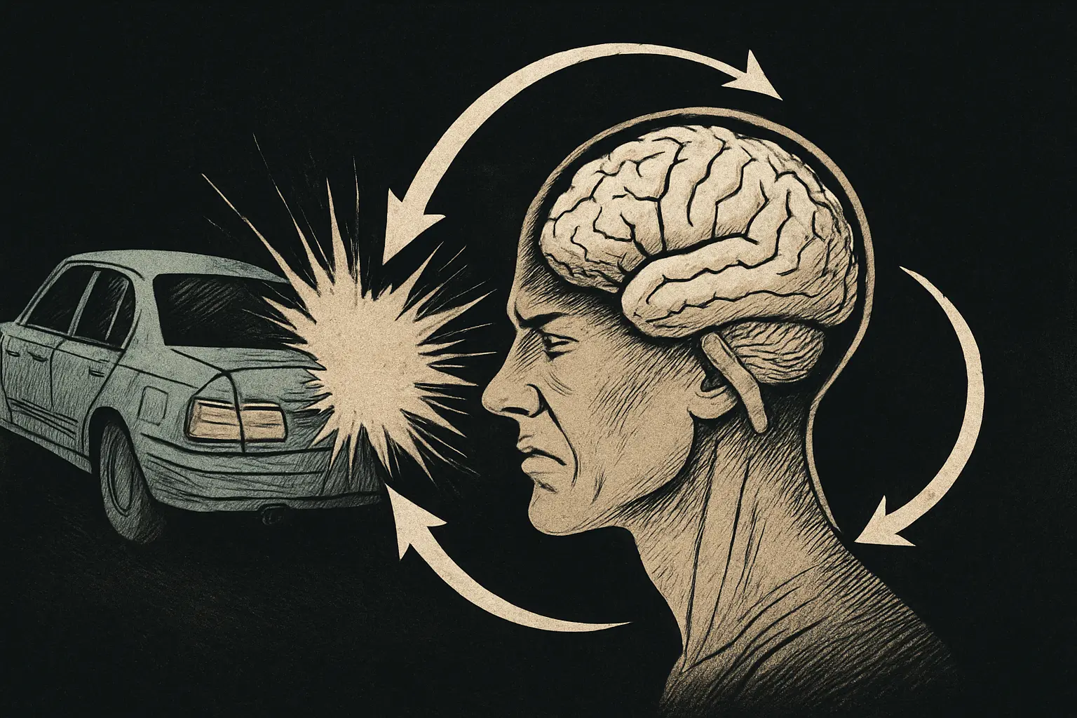
Whiplash injuries are often associated with neck pain and musculoskeletal strain, but research shows that the rapid acceleration-deceleration forces can also cause subtle brain trauma. In fact, a mild traumatic brain injury (mTBI) – even without a direct head impact – can result from a whiplash mechanism (BCMJ article). Patients may develop neurological and cognitive symptoms after whiplash (e.g. headaches, concentration problems, dizziness) despite no obvious head injury. For decades, it was controversial whether whiplash could produce “physical” brain damage, since routine scans often appeared normal. Today, advanced imaging and case studies have provided evidence of microstructural white matter injuries in some whiplash patients, offering insight into pathophysiology and validation for those reporting persistent symptoms.
Mechanism of Injury: Whiplash involves a sudden acceleration–deceleration of the head and neck (such as in a rear-end car collision). This motion can impart shearing and rotational forces to the brain inside the skull. Even without the head striking an object, the brain’s soft tissue can move relative to the skull, straining or tearing delicate axons (the long fibers of white matter that connect brain regions). This type of diffuse shearing injury to axons is known as traumatic axonal injury (TAI), a form of diffuse axonal injury typically seen in concussions and more severe TBI. Animal and post-mortem studies have shown that axonal lesions from whiplash-like trauma (without direct impact) tend to occur in the peripheral areas of the frontal and temporal lobes – essentially the outer parts of the brain that experience the greatest shear forces during whiplash (BCMJ article). In experimental models, these peripheral lesions can trigger a cascade of biochemical events: damaged neurons release excitatory neurotransmitters (e.g. glutamate), leading to excitotoxicity that can injure neighboring neurons not initially harmed. This process helps explain how a diffuse axonal injury can expand beyond the immediately torn axons, potentially causing a wider network dysfunction over time.
Primary vs Secondary Axonal Injury: Importantly, researchers distinguish between primary axonal injury, which occurs at the moment of trauma (axons snapping or stretching from the mechanical forces), and secondary axonal injury, which develops over hours to days following the trauma. In whiplash-related TAI, secondary mechanisms like impaired axonal transport, progressive swelling, and delayed disconnection of axons may lead to a delayed onset or worsening of symptoms in some patients (Frontiers Neurology, 2018). In one report, over half of documented whiplash TAI cases showed symptoms that emerged or significantly worsened days to weeks after the accident, consistent with secondary axonal degeneration rather than immediate rupture (Frontiers Neurology, 2018). For example, a patient might initially walk away from a collision with only mild issues, but develop marked neurological deficits a month later as injured axons gradually disconnect.
Injury Severity and Loss of Consciousness: Clinically, the severity of the acceleration forces (and any rotational component) influences the degree of brain injury. Strong rotational acceleration is thought to be a key factor that can even produce brief loss of consciousness (LOC) in some whiplash cases (BCMJ article). Generally, patients who suffer a concussion-level whiplash may report feeling dazed or have a momentary LOC, but many whiplash-associated brain injuries are mild enough that no LOC or only transient confusion occurs. In the past, the absence of LOC or visible trauma led some experts to assume no brain injury occurred. We now know it is possible to have microstructural white matter damage without any LOC, especially in mild TBI. For instance, a documented case involved a 14-year-old girl in a rear-end collision who had no loss of consciousness and a Glasgow Coma Scale of 15 (normal) immediately after the whiplash – yet over the next month she developed gait and motor problems, and advanced imaging eventually revealed a white matter tract injury that explained her symptoms (Frontiers Neurology, 2018).
Functional and Metabolic Changes: Beyond structural axonal injury, whiplash trauma can lead to functional changes in the brain. Imaging studies in the 1990s and 2000s using PET and SPECT found some whiplash patients (especially those with chronic symptoms) showed altered cerebral blood flow and glucose metabolism in certain brain regions. For example, some chronic whiplash patients exhibited reduced perfusion in the parieto-occipital areas of the brain on SPECT scans (BCMJ article). Initially, such findings were interpreted as evidence of subtle brain damage, though later analysis questioned whether those particular regions (parietal-occipital) correlated with the patients’ cognitive complaints. Nonetheless, more recent functional neuroimaging research continues to support central nervous system involvement: a 2016 study reported altered regional cerebral blood flow in chronic whiplash-associated disorder, suggesting abnormal brain function in these patients’ pain and sensorimotor networks (EBioMedicine, 2016) (note: reference from summary, actual study by Vállez García et al., 2016). Additionally, some whiplash sufferers develop symptoms overlapping with post-concussion syndrome (e.g. memory issues, attention deficits), implying a shared pathophysiology between whiplash and mild TBI [Neurochirurgie, 2021] (Gil & Decq, 2021 – systematic review on whiplash vs mTBI).
One of the most significant advances in understanding whiplash-related brain injury has been the use of Diffusion Tensor Imaging (DTI), an MRI-based technique sensitive to the microstructure of white matter. Conventional MRI and CT scans usually appear normal in whiplash patients because they can’t resolve microscopic diffuse axonal injuries. DTI, however, measures the diffusion of water along axons, allowing detection of subtle changes like axonal swelling, tears, or loss of coherence in fiber tracts. Since the mid-2000s, DTI studies have revealed that some whiplash patients – especially those with persistent neurological symptoms – do show objective abnormalities in white matter tracts that were previously invisible.
A 2018 review in Frontiers in Neurology highlighted that, since DTI’s introduction, at least six published case studieshave documented traumatic axonal injuries in whiplash patients despite normal CT/MRI scans (Frontiers Neurology, 2018). In these cases, DTI-based tractography (3D reconstruction of white matter fibers) showed clear evidence of tract disruption – such as fibers that appeared thinned, torn, or discontinuous – correlating with the patient’s deficits. Commonly reported DTI findings include reduced fractional anisotropy (FA) (a measure of directional water flow) or reduced fiber counts in specific tracts, indicating microstructural damage. Notably, the corpus callosum, a frequent site of diffuse axonal injury in trauma, has been shown in mild TBI to have DTI abnormalities; however, in whiplash, the tract-specific injuries can vary depending on the nature of the force and the patient’s symptoms.
Criteria for Identifying TAI in Whiplash: Clinicians and researchers have proposed diagnostic criteria to strengthen the case that DTI findings indeed represent traumatic axonal injury from whiplash (as opposed to normal anatomical variation). One suggested approach is as follows (Frontiers Neurology, 2018):
When all these factors align in an individual case, it strongly supports that the whiplash caused a mild traumatic brain injury via axonal damage in that tract.
Example DTI Findings: To illustrate, Figure 1 from Jang & Lee (2017) shows a whiplash patient’s DTI tractography with multiple injuries (Frontiers Neurology, 2018). In that case, the patient had severe symptoms (tremors, balance problems, weakness) and DTI revealed discontinuous or thinned fibers in several tracts: the corticospinal tracts (CST) (motor pathways) appeared partially torn at the subcortical level, the spinothalamic tracts (STT) (sensory pain pathways) were markedly thinner, and the corticoreticulospinal tracts (CRT) (involved in posture and locomotion) were disrupted – all despite a normal conventional MRI. This pattern of widespread axonal injury explained the patient’s multi-faceted neurologic impairments.
Group Studies: While early evidence came from single-patient case reports, more recently larger studies have begun to confirm microstructural changes in cohorts of whiplash patients. For instance, a 2023 study examined 19 patients who developed chronic central neuropathic pain after whiplash, compared to 19 healthy controls (J Integrative Neuroscience, 2023). Using DTI tractography, researchers focused on the spinothalamic tract (STT) – the pathway that transmits pain signals to the brain – because these patients had intractable pain with no peripheral cause. They found that whiplash patients had significantly reduced STT tract volume (fewer reconstructed fibers) compared to controls, consistent with structural damage or loss of some axons in the STT. Interestingly, the fractional anisotropy (FA) of the tract did not differ between groups, suggesting that standard diffusion metrics might miss certain injuries whereas tractography fiber counting captured it. Moreover, this study noted that collision mechanics mattered: patients whose whiplash came from a rear-end collision developed central pain much faster (within days) and with higher pain intensity than those in frontal-impact collisions, where pain onset was delayed (~2 weeks on average) (J Integrative Neuroscience, 2023). This hints that rear impacts (causing hyperextension of the neck) may inflict a different pattern or severity of axonal strain than frontal impacts, an observation that could guide future research on injury prevention and prognosis.
Modern imaging evidence strongly indicates that whiplash can cause microstructural white matter injuries in at least a subset of patients. These injuries, while “invisible” to CT/MRI, can be detected via DTI and provide a biological explanation for post-whiplash syndromes. In the next section, we will look at specific case examples of such injuries and the neurological consequences documented, before discussing the important legal implications of these findings for personal injury cases.
Clinical Case Examples
Case 1 – Adolescent with delayed neurological deficits:
A 14-year-old girl experienced a rear-end collision leading to a whiplash injury. Initially, she had no loss of consciousness, normal MRI, and normal neurological exam. However, over the next month, she developed progressive motor symptoms, gait disturbance, and tremors. Advanced DTI tractography revealed multiple white matter tract injuries: partial tearing of the corticospinal tracts, thinning of the spinothalamic tracts, and disruption of corticoreticulospinal tracts. These findings correlated with her neurological deficits, validating the diagnosis of whiplash-induced traumatic axonal injury (Frontiers Neurology, 2018).
Case 2 – Chronic pain syndrome after rear-end collision:
In a study of 19 patients who developed chronic central neuropathic pain after whiplash, DTI showed reduced spinothalamic tract volume compared to controls, consistent with axonal loss (J Integrative Neuroscience, 2023). Patients injured in rear-end collisions developed pain faster (within days) and more severely than those from frontal collisions, where pain onset was delayed. This suggests a link between collision mechanics, axonal injury patterns, and clinical outcomes.
Case 3 – Cognitive and psychiatric sequelae:
Some whiplash patients present not only with pain and motor deficits, but also with cognitive impairment, memory problems, and emotional dysregulation. DTI studies have revealed white matter abnormalities in frontal lobe tracts and corpus callosum in these patients, aligning with the executive dysfunction and mood changes they report (Neurochirurgie, 2021). Such cases highlight that whiplash can mimic post-concussion syndrome even without direct head trauma.
Legal and Medico-Legal Implications
The question of whether whiplash can cause brain injury has profound implications in personal injury litigation. For years, defense experts argued that whiplash without head strike could not cause brain damage, citing normal CT/MRI scans as “proof.” However, DTI and other advanced imaging now demonstrate objective microstructural injury in select patients. This evidence can be pivotal for attorneys handling cases where clients have persistent neurological or cognitive symptoms.
Key points for attorneys:
- Routine imaging is often normal: CT and MRI are insufficient to rule out traumatic axonal injury. Courts should recognize that normal scans do not preclude real injury.
- DTI provides objective evidence: Tractography demonstrating torn or thinned fibers in correlation with symptoms offers strong medico-legal documentation.
- Symptom–tract correlation matters: Establishing that the damaged tract explains the patient’s deficits (e.g., corticospinal tract and limb weakness) is critical to causation arguments.
- Rear-end collisions are high-risk: Evidence shows rear impacts more often produce rapid and severe symptoms, supporting causation in common crash scenarios.
- Secondary injury explains delayed symptoms: Attorneys can argue that worsening over weeks is consistent with secondary axonal degeneration, not malingering.
For clinicians testifying in court, citing peer-reviewed evidence that whiplash can produce white matter injury provides a scientific foundation for patient complaints. The literature base includes documented DTI case studies, group analyses, and reviews in respected journals like Frontiers in Neurology and Neurochirurgie. Presenting this data can help jurors and judges understand why a “minor” crash may lead to lasting neurological dysfunction.
Patient Education and Empowerment
For patients, learning that their symptoms have a biological basis is validating. Being told “your MRI is normal, so nothing is wrong” is frustrating and dismissive. By explaining that microstructural injuries do not show on standard imaging, providers can restore confidence and encourage adherence to rehabilitation. Patients may also benefit from knowing that advanced imaging exists, and that their complaints are not “all in their head” – rather, they reflect real though subtle brain injuries.
Conclusion
Whiplash injuries extend beyond the neck. The same forces that strain cervical ligaments can also impose shearing stress on the brain’s white matter, producing traumatic axonal injuries invisible to standard scans. Advanced imaging, particularly diffusion tensor imaging (DTI), has demonstrated these injuries in whiplash patients with persistent symptoms. Clinical cases show that white matter tract disruptions align with neurological deficits, including motor problems, pain syndromes, and cognitive changes. These findings not only advance medical understanding but also carry significant legal implications, providing objective evidence that can substantiate patient claims in court. For clinicians, attorneys, and patients alike, recognizing the neurological dimension of whiplash reframes it from a “soft-tissue neck injury” to a potential brain injury with lasting consequences.

