What two neuroscientists at Parker Seminars 2026 taught me about the single biggest thing you can do for your brain — and the one thing that will destroy it.
The Moment That Changed How I Think About Brain Injury
I've been treating personal injury patients in Petaluma for over 25 years. More than a thousand rear-end collision cases in the past four years alone. I've read the MRIs. I've documented the headaches, the cognitive fog, the sleep that never comes. I've written the reports and negotiated the liens.
But last week, sitting in two back-to-back lectures at Parker Seminars 2026 in Las Vegas, I realized I'd been thinking about brain injury recovery — and brain health in general — with only half the picture.
The first lecture was by Tommy Wood, MD, PhD — a neonatal neuroscientist from the University of Washington, performance consultant to Formula One drivers, and author of the upcoming book The Stimulated Mind. The second was by Kent Werner, MD, PhD — an active-duty Navy neurologist running one of the most advanced traumatic brain injury clinics in the military.
Wood talked about what the brain needs. Werner talked about what happens when it doesn't get it.
Together, they painted a picture I can't unsee — and one I believe every patient recovering from an injury, every person worried about cognitive decline, and every clinician managing these cases needs to understand.
Here's what I learned.
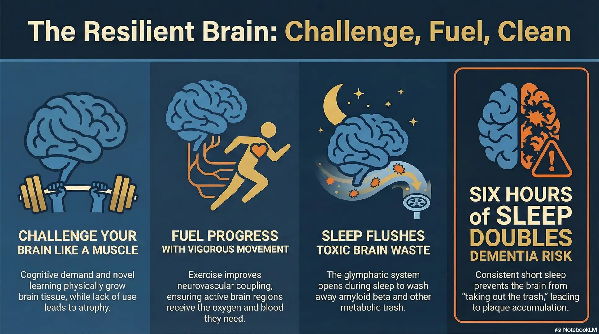
Part I: Your Brain Is a Muscle — And It's Begging to Be Worked
I know you've probably heard someone say the brain is "like a muscle." It sounds like a bumper sticker. But Wood presented a body of evidence that makes the analogy almost literal.
His model is elegant: Stimulus → Supply → Support.
Your brain requires demand — cognitive challenge, novel learning, social interaction — the way your biceps require resistance. Without stimulus, the tissue atrophies. With it, the tissue remodels and grows. This isn't metaphor. It's measurable on MRI.

The London Taxi Driver Study
Consider one of the most famous neuroplasticity studies ever conducted. Eleanor Maguire and Katherine Woollett at University College London followed aspiring London taxi drivers as they spent three to four years memorizing 25,000 streets within a six-mile radius of Charing Cross Station — a grueling process called "The Knowledge."
The drivers who passed the exam showed significant structural growth in their posterior hippocampus — the brain region responsible for spatial memory. Those who failed showed no changes. And here's the trade-off that fascinated Wood: the successful drivers became worse at abstract visual memory tasks. The brain didn't just grow — it reorganized. It adapted to exactly what was demanded of it, and nothing more.
Key Takeaway
The brain doesn't just grow in response to learning — it reorganizes. It adapts to exactly what you demand of it. Taxi drivers gained spatial memory but lost abstract visual memory. Use it or lose it is literal.
The Juggling Study
Another landmark: Bogdan Draganski's team taught adults in their twenties and thirties to juggle a three-ball cascade over three months. MRI revealed gray matter increases in the visual cortex areas associated with processing complex motion. Better jugglers showed bigger changes. But when they stopped juggling for three months, the structural gains began reversing.
Use it or lose it — even in young, healthy adults.
Processing Speed: The 20-Year Evidence
Wood's most practice-changing evidence came from the ACTIVE Study — Advanced Cognitive Training for Independent and Vital Elderly. Starting in 1998, nearly 3,000 adults aged 65 to 94 were randomized into four groups: control, memory training, reasoning training, and processing speed training.
The processing speed group used a computerized task — now available as "Double Decision" on BrainHQ — that requires identifying objects in the center of a screen while simultaneously locating peripheral targets, all at increasingly brief exposures.
The results were staggering:
- At five years, the speed training group showed better health-related quality of life, was more likely to still be driving, and had fewer car accidents.
- At ten years, both reasoning and speed training showed sustained cognitive improvement.
- At twenty years — follow-up data published just weeks before the seminar — speed training with booster sessions showed a statistically significant reduction in dementia risk.
20 yrs
of follow-up data showing processing speed training reduces dementia risk
No supplement. No drug. A computerized visual processing task, practiced consistently, reduced the likelihood of dementia two decades later.
A subsequent neuroimaging study of about 100 individuals in their seventies found that speed training improved cholinergic signaling in the anterior cingulate cortex — a region critical for attention, executive function, and decision-making. Cholinergic decline is a hallmark of aging-related cognitive loss. The training was essentially reversing one of the neurochemical signatures of brain aging.
You can't just take naps and drink protein shakes. You actually have to lift the weights. And the brain is essentially the same. — Tommy Wood, MD, PhD
Part II: The Brain's Cleaning Crew Works the Night Shift
If Wood's lecture was about what the brain needs during waking hours, Werner's was about what happens during the hours most of us take for granted.
And it starts with a system most clinicians have never heard of.

The Glymphatic System
In 2013, Maiken Nedergaard's lab at the University of Rochester published a landmark paper in Science that redefined our understanding of sleep. Using two-photon imaging in live mice, they demonstrated that during sleep, the interstitial spaces between brain cells expand by approximately 60%, creating a convective highway for cerebrospinal fluid to flush through brain tissue.
This system — dubbed "glymphatic" because glial cells facilitate lymphatic-like drainage in an organ that has no true lymph vessels — clears metabolic waste products including amyloid beta and tau protein, the molecular hallmarks of Alzheimer's disease.
The name is new. The implication is ancient: sleep is when the brain takes out its trash.
The mechanism is elegant. Aquaporin-4 (AQP4) water channels on astrocyte endfeet migrate to the blood-brain barrier interface during sleep, allowing CSF to flow along perivascular spaces — the Virchow-Robin spaces — deep into brain tissue. During wakefulness, sympathetic nervous system activity constricts these channels. During sleep, the parasympathetic system opens them.
Important
Sympathetic activation — the fight-or-flight response — completely blocks glymphatic clearance. A trauma patient who lies hypervigilant at night isn't just sleeping poorly. The brain is physically unable to clear the toxic byproducts of injury.
Part III: When Sleep Fails, the Brain Pays
I know you might think that six hours of sleep is "enough." Many of my patients tell me exactly that. Some wear it as a badge of honor.
Here's what the data actually shows.

Six Hours or Less Doubles Dementia Risk
Werner cited a study following 4,000 people for 30 years using wrist actigraphy — a wearable device that measures movement patterns to objectively estimate sleep duration and quality. Those sleeping six hours or less had twice the rate of dementia compared to those getting seven to eight hours.
The relationship was U-shaped — long sleepers (more than nine hours) also showed elevated risk, but Werner's interpretation was pointed: excessive sleep isn't the problem. It's a red flag for undiagnosed sleep quality problems — untreated apnea, circadian dysfunction, or other disorders masquerading as a need for more time in bed.
Sleep Apnea and the 70% Reduction
Kristine Yaffe's group at UCSF followed 107 women in their seventies for five years. Those with sleep-disordered breathing were nearly twice as likely to develop mild cognitive impairment or dementia. But those who treated their sleep apnea — bringing oxygen desaturation events below 15 per hour — showed approximately 70% less dementia.
70%
reduction in dementia from treating sleep apnea alone
Amyloid Accumulates in the Short Sleepers
Adam Spira's team at Johns Hopkins used Pittsburgh Compound B (PiB) PET imaging to scan the brains of healthy people in their fifties — no cognitive complaints, no diagnosis. Those who self-reported sleeping six hours or less had significantly greater amyloid plaque burden than those who slept longer.
Amyloid beta is a normal byproduct of neuronal activity. You produce it every time you think. The glymphatic system clears it during sleep. When you cut sleep short, the waste accumulates. Night after night, year after year.
Part IV: TBI Breaks the System — And Sleep Is Ground Zero
This is where the two lectures converged for me, and where the implications for my practice became impossible to ignore.
The Feed-Forward Cycle
Werner's central hypothesis — supported by his own lab's unpublished data and a growing body of published research — is devastatingly simple:
TBI → disrupted sleep-wake circuitry → impaired glymphatic clearance + elevated sympathetic tone → waste accumulation + chronic inflammation → accelerated neurodegeneration → which further disrupts sleep → repeat.
It's a vicious cycle. And the intervention point is sleep.
Jeffrey Iliff's group demonstrated this in rodent models: head injury reduces glymphatic flow by day one, and it worsens by day seven — even on the uninjured side of the brain. The mechanism isn't purely structural. It's the systemic sympathetic activation that follows trauma, disrupting the parasympathetic state required for glymphatic function.

200,000 Veterans, 14 Years of Follow-Up
Yue Leng and Kristine Yaffe's group studied nearly 200,000 US veterans over 14 years. Those with TBI had a 40% greater incidence of sleep disorders — insomnia, sleep apnea, hypersomnia, and movement disorders — even after controlling for PTSD, medications, and substance use.
Werner added a clinical observation from his own TBI clinic: 50% of young special operators referred for cognitive complaints end up diagnosed with obstructive sleep apnea. Not the classic profile — not obese, not elderly. Young, fit, brain-injured soldiers whose arousal thresholds have been permanently altered by trauma.
Blood Biomarkers Tell the Story Years Later
Perhaps most disturbing: blood samples from veterans 8 to 10 years after mild TBI show elevated amyloid beta, exosomal tau, and neurofilament light chain (NfL) — markers of ongoing neurodegeneration. And among those TBI patients, the poor sleepers had the highest levels of NfL and inflammatory markers like IL-6 and TNF-alpha.
The injury happened a decade ago. The brain is still deteriorating. And the difference between progressive decline and stabilization may come down to sleep quality.
Part V: What Actually Works — The Practical Toolkit
Both Wood and Werner are clinician-scientists. They don't just study the problem — they treat patients. Here's what they recommend, synthesized from both lectures.
Stimulus: Challenge Your Brain Daily
Wood's demand-function coupling model argues that cognitive demand is the primary upstream driver of long-term brain health. The evidence supports:
- Processing speed training via BrainHQ "Double Decision" — the only cognitive training with 20-year dementia prevention data
- Complex real-world skills — music, dance, art, language learning. A 2024 meta-analysis found that greater expertise in creative disciplines correlated with younger-looking brain network connectivity
- Socially demanding work — jobs requiring complex problem-solving and interpersonal interaction show lower dementia risk, even in people with low educational attainment
- Avoid cognitive prosthetics — an MIT study ("Your Brain on ChatGPT") showed that students using AI for essay writing had up to 55% reduced brain connectivity compared to those who wrote unaided, with 83% unable to recall content from essays they had just "written"
Did You Know
Students who wrote essays themselves first and then used AI to extend their work produced significantly better results — because they engaged their brains in the process. The key question: Is this tool augmenting my capabilities, or replacing important skills?
Supply: Protect Cardiovascular and Metabolic Health
Wood's model emphasizes that stimulus is useless without adequate blood flow, oxygen, and metabolic substrates reaching active brain tissue:
- Neurovascular coupling draws blood to active brain regions — but only if cardiovascular health supports it
- Exercise is dual-purpose: it improves supply (blood flow, BDNF release) and builds cross-stress adaptation. People who exercise regularly show smaller emotional disruptions under social stress
- 20 minutes of vigorous exercise improves cognition for one to two hours afterward — a practical tool before demanding clinical or intellectual work
Support: Protect Sleep as If Your Brain Depends on It — Because It Does
This is where Werner's clinical expertise shines. His practical sleep optimization protocol:
Environment
- Bedroom temperature: 65°F (REM sleep requires thermoregulatory passivity — the body stops regulating temperature during REM, so the room must do it)
- Pitch darkness — Phyllis Zee's lab at Northwestern found that even ambient light from a Wi-Fi router or alarm clock predicted higher BMI and disrupted circadian rhythm. The standard: you shouldn't be able to see your hand in front of your face.
- No food within 3 hours of bedtime — Zee's research showed that eating one hour before sleep elevated inflammation, insulin insensitivity, and heart rate. The same meal three hours before sleep normalized all markers.
Supplementation
- Magnesium oxide — Werner's number one sleep-enhancing agent. Study protocol: 1,200 mg at night + 1,200 mg in the morning. Significantly deeper sleep on EEG plus better cognitive performance. Mechanism: magnesium is nature's endogenous neuronal inhibitor.
- Melatonin 0.5 mg of a third-party tested brand — meta-analysis shows benefit specifically in TBI patients. Take at least one hour away from food.
Behavioral Interventions
- Cognitive Behavioral Therapy for Insomnia (CBTI) — best long-term outcomes for those who complete it. Free resources: Insomnia Coach app (VA) and Path to Better Sleep (VA website)
- Morning bright light exposure — 10,000+ lux speaks to intrinsically photosensitive retinal ganglion cells, directly regulating the circadian clock. Outdoor sunlight delivers approximately 100,000 lux even on cloudy days.
Medications to Consider (Under Clinical Supervision)
- Orexin antagonists (suvorexant, lemborexant, daridorexant/Quviviq) — unlike Ambien and Lunesta, which may block glymphatic clearance, orexin antagonists have been shown in rodent models to preserve glymphatic activity and even prevent Alzheimer's pathology
- Prazosin for trauma-related sleep disruption — an alpha blocker shown to enhance glymphatic activity in rodent models, often dramatically effective for dream enactment and nightmares in PTSD patients
Manage Your Stress Response
Wood introduced Alia Crum's research at Stanford on stress mindset. When participants were trained to view stress as enhancing rather than debilitating before undergoing acute stress testing, both groups showed cortisol spikes — but the "stress-is-enhancing" group also spiked DHEA, a neurosteroid that promotes functional adaptation. The higher the DHEA-to-cortisol ratio, the better the decision-making under pressure.
The clinical translation: how you frame the stress of recovery matters. Not toxic positivity. Not denial. A genuine reorientation toward the idea that the discomfort of rehabilitation is the stimulus your brain needs to rebuild.
Part VI: What This Means for You
If you're recovering from a car accident, a fall, a concussion — or if you're simply someone who wants to protect the organ that makes you you — here's the picture these two scientists painted:
Your brain is not a passive organ waiting for damage to accumulate. It is an active, adaptive, structurally dynamic system that responds to what you demand of it. Challenge it, and it grows. Neglect it, and it withers. Injure it, and it enters a cycle that can be interrupted — but only if you intervene at the right points.
The right points are:
- Stimulus — Engage in cognitively demanding activities every day. Learn something difficult. Have complex conversations. Practice processing speed. Don't outsource your thinking to machines.
- Supply — Exercise. Protect your cardiovascular health. Give your brain the blood flow it needs to respond to demand.
- Support — Sleep seven to eight hours in a dark, cool room. Address sleep apnea aggressively. Use evidence-based supplements. Avoid eating close to bedtime. And if you've had a brain injury, treat sleep optimization as the primary therapeutic intervention — because the glymphatic system that clears the toxic aftermath of your injury only operates when you're asleep.
Wood ended his lecture with a quote about self-compassion — that the strongest predictor of long-term success in elite athletes isn't grit or punishment, but the ability to treat yourself with the same patience you'd offer someone you care about.
Werner ended his with a hypothesis and a promise: that the feed-forward cycle of TBI and neurodegeneration can be broken, and that the tools to break it — from focused ultrasound to non-invasive brain stimulation to simple magnesium supplementation — either exist now or are arriving within years.
"Every patient who walks into my clinic after a rear-end collision isn't just dealing with neck pain and headaches. They're standing at the entrance to a cycle that, left unmanaged, could reshape the trajectory of their cognitive life."
— Dr. Todd Lloyd, D.C.
The most powerful thing I can do — before the MRI, before the report, before the lien negotiation — is make sure they understand what their brain needs to heal.
It needs to be challenged. It needs to be fed. And above all, it needs to sleep.
References
- Xie L, Kang H, Xu Q, et al. Sleep drives metabolite clearance from the adult brain. Science. 2013;342(6156):373-377. PubMed
- Iliff JJ, Chen MJ, Plog BA, et al. Impairment of glymphatic pathway function promotes tau pathology after traumatic brain injury. J Neurosci. 2014;34(49):16180-16193. PubMed
- Maguire EA, Woollett K, Spiers HJ. London taxi drivers and bus drivers: a structural MRI and neuropsychological analysis. Hippocampus. 2006;16(12):1091-1101. PubMed
- Woollett K, Maguire EA. Acquiring "the Knowledge" of London's layout drives structural brain changes. Curr Biol. 2011;21(24):2109-2114. PubMed
- Draganski B, Gaser C, Busch V, et al. Neuroplasticity: changes in grey matter induced by training. Nature. 2004;427(6972):311-312. PubMed
- Ball K, Berch DB, Helmers KF, et al. Effects of cognitive training interventions with older adults: a randomized controlled trial. JAMA. 2002;288(18):2271-2281. PubMed
- Willis SL, Tennstedt SL, Marsiske M, et al. Long-term effects of cognitive training on everyday functional outcomes in older adults. JAMA. 2006;296(23):2805-2814. PubMed
- Yaffe K, Laffan AM, Harrison SL, et al. Sleep-disordered breathing, hypoxia, and risk of mild cognitive impairment and dementia in older women. JAMA. 2011;306(6):613-619. PubMed
- Spira AP, Gamaldo AA, An Y, et al. Self-reported sleep and β-amyloid deposition in community-dwelling older adults. JAMA Neurol. 2013;70(12):1537-1543. PubMed
- Leng Y, Byers AL, Barnes DE, et al. Traumatic brain injury and incidence risk of sleep disorders in nearly 200,000 US veterans. Neurology. 2021;96(13):e1792-e1799. PubMed
- Mysliwiec V, O'Reilly B, Polchinski J, et al. Trauma associated sleep disorder: a parasomnia induced by trauma. Sleep Med Rev. 2018;37:94-104. PubMed
- Crum AJ, Salovey P, Achor S. Rethinking stress: the role of mindsets in determining the stress response. J Pers Soc Psychol. 2013;104(4):716-733. PubMed
- Wood T, Turley J. Is the lack of appropriate cognitive demand the primary driver of dementia risk? Lifestyle Med. 2022;3(4):e70. Wiley
- Kosmyna N, et al. Your brain on ChatGPT: Accumulation of cognitive debt when using an AI assistant for essay writing task. arXiv preprint arXiv:2506.08872. 2025. MIT Media Lab
- Jessen NA, Munk AS, Lundgaard I, Nedergaard M. The glymphatic system: a beginner's guide. Neurochem Res. 2015;40(12):2583-2599. PMC
Based on lectures by Tommy Wood, MD, PhD and Kent Werner, MD, PhD at Parker Seminars 2026, Las Vegas. Essay by Todd Lloyd, DC — ADJUST.CLINIC, Petaluma, CA.